Pesquisar neste blogue

quinta-feira, 23 de fevereiro de 2017

5 Steps to Flexibility

Fonte: amnacademy.com

What is Flexibility?

At AMN, we define flexibility as strength at end ranges of motion. In a more holistic sense, however, flexibility is freedom.
The more strength we have at end ranges of motion, the more at ease our bodies can adapt to new movements – we can ultimately perform existing movement skills to greater quality. It allows us to explore extended positions of the limbs, allows us to flow effortlessly from one position to another; all of this with a reduced need for strength… whilst looking bendy and efficient.
When we lack flexibility, it feels as though our bodies fight back at us – Our minds are saying: ‘There is no way I can do that!’ We are so ingrained in our little movement box, that when faced with a movement task that ventures astray from the typical small range of motion, we put serious strain on our body. In the end, we hurt ourselves.
Truth be said, flexibility is a poorly understood and poorly executed pursuit within the health and fitness industry. This has resulted in it being demoted from an important primary physical attribute, to the cursory towel biting hamstring stretch at the end of a training session.
When we look outside of the health and fitness biz, we find a lot of flexible people in dance, gymnastics and martial arts. There is a huge amount to learn from the way these practices approach movement and flexibility training… Unfortunately, for most of us, it is hard to dedicate 6+ hours per week that a gymnast may dedicate to flexibility work.
AT AMN, WE DEFINE FLEXIBILITY AS STRENGTH AT END RANGES OF MOTION. IN A MORE HOLISTIC SENSE, HOWEVER, FLEXIBILITY IS FREEDOM.
What we’ve done at AMN is taken inspiration from these disciplines, and paired them with a neurological rationale… Ultimately producing a five-step process to gaining flexibility. This is for the typical personal training client… The type of client that you and I work with.
This is also the process that I utilise to improve my flexibility – because let me tell you – I am not the naturally bendy type. I also skipped the official dance and gymnastics training as a child, that I now see serves my colleagues so well throughout adulthood.

The Five Steps Process to Gaining Flexibility

Step One – Define a position specific goal

This is a very important part of the process. It defines where your starting point is and what the end goal should be, and therefore serves as a bench mark of progress.
While there are a whole host of positions you could go for, we chose the pike fold, the straddle pancake, the bridge, and the squat as the four primary specific positions to work towards. These may vary depending on your goal; for the purposes of AMN movement training, these positions work synergistically with gymnastics strength – skills we promote in our Fundamentals Pro exercise program, with movements such as handstands, straddle positions, skin the cat etc.

Step Two – Joint mobility sequencing

Here’s where the first neuro-rationale comes in. A joint mobility sequence is a single or series of movements designed to reduce levels of resting tension in the muscles. We take this step as we deal with adults… Tight, inflexible adults.
When we perform a joint mobility sequence, we utilise novel and complex movement patterns that are proprioceptively stimulating to the brain. When it comes to movement, a stimulated brain is a brain which is paying attention.
The end-result of practicing novel and complex movement with minimal tension: improved muscle recruitment and reduced muscular tension, local to the drill and elsewhere in the body.
This prepares the system to be more responsive to stretching protocols.

Step Three – Contract-Relax stretching

Contract-relax stretching takes the muscles in to a lengthened position and then contracts the stretching muscle against resistance.
This process increases tension in the muscle while stretched, and stimulates the Golgi-tendon organs (GTO’s). The GTO’s are part of the neuromuscular system which are sensitive to tension in the muscle tissues. When we stimulate them with contract-relax techniques, we reduce the sensitivity of the muscle spindles to stretch and relax the alpha motor neurons, which are the other parts of the neuromuscular system governing your current ranges of motion.
WHEN IT COMES TO MOVEMENT, A STIMULATED BRAIN IS A BRAIN WHICH IS PAYING ATTENTION.
The net result is the ability to move the joint in to a new range of motion, in which the process may be repeated. When it comes to gaining strength at end range of motion, flexibility and contract-relax protocols form an imperative part of the peripheral nervous systems (PNS) education.
The PNS must learn how to take the brakes off the muscles when we lengthen them. Thus, we need to speak the right language in an effort to communicate the right message. Learning to contract and relax in lengthened positions forms a part of this dialogue.

Step Four – Loaded stretching

Now back to the bit about strength at end range of motion – Let me paint you a picture: If you can get your foot in your mouth… and then on top of this have the ability to contract your glutes… that’s strength at end range of motion!
Strength at end range of motion is definitely a skill that can be learnt and trained.
When we can contract appropriately in lengthened positions, we have control of the tissues and joints. This equates to trust between the nervous system and the muscles. Trust results in reduced tension, and fight from your own body. After that, the funky contortions are endless…
Loaded stretching further increases stimulation to the GTO’s through dynamic ranges of motion in full stretch positions. It is different to contract-relax stretching as it is void of the complete relaxation phase.
Tension is increased though the addition of external load and controlled as the body is taken through increasing ranges of stretch. Once an external load is incorporated and movement utilised, load sharing between the agonist and antagonist muscles is completely altered – as is the demand on core activation.
WHEN IT COMES TO GAINING STRENGTH AT END RANGE OF MOTION, FLEXIBILITY AND CONTRACT-RELAX PROTOCOLS FORM AN IMPERATIVE PART OF THE PERIPHERAL NERVOUS SYSTEMS (PNS) EDUCATION.
In this part of the process – Strength can be developed, and increased range of motion realised at a faster rate, in comparison to with no external loading. It also allows us to utilise load and volume progressions, which is incredibly useful feedback for progress.
david-fleming-fundamentals-stretch

Step Five – Integrated movements

Here’s the bit about freedom…
Having worked on reducing overall tension with mobility sequences, re-educating the sensitivity of the neuromuscular system to stretch with contract-relax stretching, and increasing strength in new ranges with loaded stretching, Integrated movements provide context in which to utilise new flexibility.
When context is provided to movement, it tends to improve. This is the difference between working on the parts and integrating the whole. For example, if you ask someone to lie down and show you their active straight leg raise range, and then compare that range to what is achieved if you provide them with a target to reach towards, they will inevitably perform better when the movement has context.
As a movement is practiced, less attention is placed on the specifics of how tight something feels. Integrated movements are an expression of flexibility – this is important in demonstrating to the client that they are very well capable of moving with increased levels of freedom.
Fluidity and efficiency in movement, through increasing ranges of motion is the kind of flexibility that people want and should work towards. Static positions are useful in defining progress – but enhanced movement capabilities are where you experience the rewards of improved flexibility.
fundamentals-of-amn-david-fleming

How long does it take?

Like all health and fitness attributes, people have varying natural abilities and starting points – Flexibility is no different. With this in mind, it naturally takes some people longer than others and some will have to focus more or less on different aspects of the process above.
But, no matter what the starting point is – a neurologically sound approach to flexibility training, that is sensitive to the typical personal training client, provides gradual progress – with no wasted time on ineffective methods.
When it comes to flexibility, you get out what you put in. It is an extremely important physical attribute that should be at the forefront of the movement aspect of health and wellness programmes.
Personal trainers have an amazing opportunity to significantly impact their clients’ strength and flexibility progress. I know that many of my current and past clients have been absolutely shocked at the amount of progress they have made in these two departments… If you can make your client achieve something that they’ve never before thought was ever possible… it’s a pretty good feeling. Especially when it comes to improving flexibility – something many people think you are either born with… or without!
FLUIDITY AND EFFICIENCY IN MOVEMENT, THROUGH INCREASING RANGES OF MOTION IS THE KIND OF FLEXIBILITY THAT PEOPLE WANT AND SHOULD WORK TOWARDS.

Our Fundamentals Pro course is a comprehensive guide to Strength & Flexibility with an emphasis on gymnastics based skills.
Created specifically for Personal Trainers that want to expand their knowledge in these fields, and differentiate themselves from the norm; this course contains over 100 video tutorials, a comprehensive manual delving into neuroanatomy, program design and gives an in-depth understanding of movement and how that can positively affect the Brain and Nervous System.

segunda-feira, 20 de fevereiro de 2017

Can these 4 things dramatically reduce your cancer death risk?

Fonte: http://www.ahchealthenews.com

Cleveland Clinic Performs Nation’s First Deep Brain Stimulation for Stroke Recovery

Fonte:https://newsroom.clevelandclinic.org

Cleveland Clinic has performed the first deep brain stimulation (DBS) surgery for stroke recovery, as part of an ongoing clinical trial assessing the procedure’s potential to improve movement in patients recovering from stroke.
Stroke is the leading cause of long-term disabilities in the United States. Despite rehabilitative efforts, one-third of stroke patients maintain long-term motor deficits severe enough to be disabling.
A team led by Andre Machado, M.D., Ph.D., chairman of Cleveland Clinic’s Neurological Institute, performed the DBS surgery Dec. 19, 2016. During the 6 hour procedure, electrodes were implanted in a part of the patient’s brain called cerebellum, which has extensive connections with the cerebral cortex. Connected to a pace-maker device, DBS electrodes provide small electric pulses as a way to help people recover control of their movements.
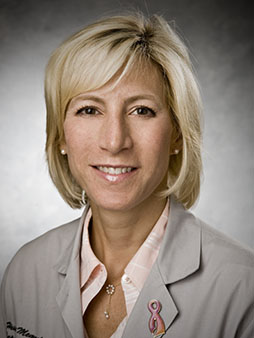
Andre Machado, M.D., Ph.D., chairman of Cleveland Clinic’s Neurological Institute
“If this research succeeds, it is a new hope for patients that have suffered a stroke and have remained paralyzed after a stroke. It is an opportunity to allow our patients to rehabilitate and gain function and therefore gain independence,” Dr. Machado said. “Our knowledge to date shows that deep brain stimulation can help the brain reorganize, can help the brain adapt, beyond what physical therapy alone can do. The goal of our study is to boost rehabilitation outcomes beyond what physical therapy alone could achieve.”
Over the next few weeks, the patient – who has been discharged home feeling well and in stable condition – will continue to heal and recover from brain surgery, followed by physical therapy. After a few weeks of rehabilitation, the DBS device will be turned on as the patient continues physical therapy. The patient will be monitored and evaluated regularly to determine how DBS can boost the effects of physical therapy.
“In addition to characterizing the effect of treatment on motor recovery, we will examine directly how stimulation affects brain activity using a combination of non-invasive imaging and electrophysiological techniques,” said Kenneth Baker, Ph.D., of Cleveland Clinic’s Department of Neurosciences and co-primary investigator on the research grant.  “Through these studies, we hope to gain further insight into its therapeutic mechanisms and, perhaps more importantly, how best to optimize delivery of the therapy as we move forward.”
Dr. Machado’s previous research has shown that DBS targeting the same brain pathway in a laboratory model promotes the brain’s plasticity, the ability to form new neural connections, during recovery from stroke. This clinical trial expands on that work and for the first time translates it to humans. This is currently an experimental approach and, as for any surgical procedure, has risks. Potential risks include hemorrhage, infection and neurological complications. Additional information about the trial can be found at https://clinicaltrials.gov/ct2/show/NCT02835443.
This first-in-human trial is co-funded by an NIH BRAIN Initiative Grant: Brain Research through Advancing Innovative Neurotechnologies and this is among one of many projects exploring human brain activity.
Dr. Machado patented the DBS method in stroke recovery. Boston Scientific owns a license to those patents and provided the Vercise DBS systems used in the trial. In 2010, Cleveland Clinic Innovations established a for-profit spin-off company, Enspire DBS Therapy to fund the clinical trial and commercialize the method; Dr. Machado holds stock options and equity ownership rights with Enspire and serves as the chief scientific officer. Boston Scientific recently invested $2.5 million into Enspire DBS.


About Cleveland Clinic
Cleveland Clinic is a nonprofit multispecialty academic medical center that integrates clinical and hospital care with research and education. Located in Cleveland, Ohio, it was founded in 1921 by four renowned physicians with a vision of providing outstanding patient care based upon the principles of cooperation, compassion and innovation. Cleveland Clinic has pioneered many medical breakthroughs, including coronary artery bypass surgery and the first face transplant in the United States. U.S.News & World Report consistently names Cleveland Clinic as one of the nation’s best hospitals in its annual “America’s Best Hospitals” survey. Among Cleveland Clinic’s 49,000 employees are more than 3,400 full-time salaried physicians and researchers and 14,000 nurses, representing 120 medical specialties and subspecialties. The Cleveland Clinic health system includes a 165-acre main campus near downtown Cleveland, nine community hospitals, more than 150 northern Ohio outpatient locations – including 18 full-service family health centers and three health and wellness centers – and locations in Weston, Fla.; Las Vegas, Nev.; Toronto, Canada; Abu Dhabi, UAE; and London, England. In 2015, there were 6.6 million outpatient visits, 164,700 hospital admissions and 208,807 surgical cases throughout the Cleveland Clinic health system. Patients came for treatment from every state and 180 countries. Visit us at www.clevelandclinic.org.  Follow us at www.twitter.com/ClevelandClinic.

quinta-feira, 9 de fevereiro de 2017

segunda-feira, 6 de fevereiro de 2017

Reabilitação física em um paciente com a Doença de Charcot-Marie-Tooth: Relato de caso

Fonte: Rev Neurocienc 2009


Autores: Talita Helen Ferreira e Vieira1 , Rosária Dias Aires1 , Vanessa Amaral Mendonça2 * Clynton Lourenço Corrêa3


RESUMO

Introdução. A doença de Charcot-Marrie-Tooth (CMT) é uma neuropatia periférica hereditária caracterizada por atrofia muscular e hipoestesia distal dos membros podendo estar associada à hipotonia, fraqueza muscular, diminuição dos reflexos profundos, fadiga e alterações respiratórias. O objetivo do estudo foi avaliar os efeitos da fisioterapia motora, do treinamento de condicionamento físico e do treino muscular respiratório em uma paciente com a doença de CMT. Método. As variáveis analisadas foram: Pressão Inspirató- ria Máxima (PImáx), Pressão Expiratória Máxima (PEmáx), Teste de Caminhada de 6 minutos, Timed up and go, os Testes de Velocidade Média da Marcha, Subida e Descida de Escadas e o Questionário de Qualidade de Vida SF-36. Resultados. Após 8 semanas de tratamento foram encontrados aumento dos valores da PImáx e PEmáx, da distância percorrida no teste de caminhada de 6 minutos, na pontuação atingida no questionário SF-36, na velocidade média da marcha e nos tempos de realização dos testes de subida e descida de escadas. Houve diminuição no tempo de realização do timed up and go.

 Conclusão. O treino de força muscular respiratória e o treinamento físico associados à fisioterapia convencional neurológica podem potencializar a funcionalidade motora e respiratória e prevenir a fadiga em pacientes com doença de CMT.

 Keywords: Condicionamento Físico Hmano. Exercícios Respiratórios. Fisioterapia. Qualidade de Vida. Doença de Charcot-Marie-Tooth. Citação: Vieira THF, Aires RD, Mendonça VA, Corrêa CL. Reabilitação física em um paciente com a Doença de CharcotMarie-Tooth: Relato de caso.

A doença de Charcot-Marie-Tooth (CMT), também conhecida como neuropatia hereditária sensitiva motora (NHSM), é a mais freqüente das doenças neuromusculares hereditárias, podendo ser classificada em diversos tipos distintos1-3. A doença de CMT é comumente classificada como tipo I (desmielinizante), em que ocorre desmielinização e grande redução na velocidade de condução nervosa ou tipo II (axonal), em que ocorre degeneração axonal primária e a velocidade de condução nervosa está normal ou ligeiramente comprometida4-6. Clinicamente a doença de CMT é caracterizada por amiotrofia, paresia, hipoestesia e arreflexia distal em membros inferiores, podendo também acometer os membros superiores1,2,4. Seus primeiros sintomas geralmente aparecem na 1a ou 2a décadas de vida2,4, com distúrbios na marcha ocasionados pela queda do pé e redução da propriocepção2 . Com a evolução da doença, o indivíduo pode apresentar outros sintomas, tais como escoliose, pé cavo e tremor nas mãos2-4,7. A maioria das causas de morbidade e mortalidade dos pacientes com doenças neuromusculares é a falência respiratória8,9. O primeiro relato de disfunção respiratória na doença CMT foi descrita por Chan et al., os quais descreveram 2 pacientes com fraqueza diafragmá- tica10,11. Posteriormente, Laroche et al., também descreveram 2 pacientes com CMT com a mesma alteração no diafragma11 e Gilchrist et al., encontraram atrofia neurogênica diafragmática e alterações no nervo frênico em autópsia de pacientes, com doença de CMT, que morreram de falência respiratória11,12. A presença da fraqueza dos músculos respiratórios associada à imobilidade, geralmente apresentada por pacientes portadores de doen- ças neuromusculares, pode levar o indivíduo a um quadro de descondicionamento13 e conseqüentemente a um importante quadro de limitações físicas. Analisando este fato e levando em considera- ção a escassez de estudos associando a fisioterapia motora, ao treino muscular respiratório e ao treinamento de condicionamento físico nos pacientes com doença de CMT, o objetivo do presente estudo destina-se a avaliar os efeitos da associação dessas três modalidades de tratamento em um paciente com essa doença.

MÉTODO

 Relato do Caso Paciente IMR, sexo feminino, 49 anos, com diagnóstico da doença de CMT 1A confirmado por exame genético há um ano, foi admitida e avaliada na Clínica-Escola de Fisioterapia da Universidade Federal dos Vales do Jequitinhonha e Mucuri (UFVJM). A paciente apresentava marcha escarvante, atrofia, fraqueza e hipotonia muscular, com predomínio em membros inferiores, hipoestesia e hiporreflexia distal, bem como escoliose e tremor nas mãos. Segundo a paciente, ela apresentava dificuldades para deambular, sentar-se em locais baixos, manter-se de pé por tempo prolongado, subir e descer escadas. Há dois anos relatou dispnéia acompanhada de fadiga ao deambular em solos inclinados.

 Procedimento 

Após entender o protocolo experimental e concordar em participar do estudo, a paciente assinou um termo de consentimento livre e esclarecido de acordo com a resolução 196/96 do Conselho Nacional de Saúde e, em seguida, foi avaliada. Foram realizadas avaliações neurológicas e respiratórias de modo a coletar dados objetivos, além da avaliação cardiorrespiratória através do Teste de Caminhada de 6 Minutos. Para a coleta de dados subjetivos foi aplicado o Questionário de Qualidade de Vida (SF- 36). Todos os procedimentos foram realizados na Clínica-Escola de Fisioterapia da UFVJM no período de outubro a dezembro de 2006. Na avaliação neurológica foram coletados dados específicos referentes a uma avaliação de lesão de nervos periféricos, como força, tônus, reflexos, sensibilidade e equilíbrio. Além disso, foram aplicados os testes Timed Up and Go, Velocidade da Marcha e Subida e Descida de Escadas. Na avaliação respiratória foram coletados dados referentes à força da musculatura respiratória através das medidas da Pressão Inspiratória Máxima (PImáx) e da Pressão Expiratória Máxima (PEmáx). Para as medidas da força da musculatura respiratória foi usado um Manovacuômetro MV-150/300 fabricante Ger-Ar calibrado e um bucal descartável. A paciente estava em posição sentada com o nariz ocluí- do por um nasoclip. Os esforços respiratórios máximos foram sustentados durante pelo menos dois segundos, sem deixar que o ar escapasse. Foi anotada a pressão mais alta alcançada após o primeiro segundo. As manobras foram repetidas por, no máximo, cinco vezes e três dessas medidas foram consideradas reprodutíveis, desde que os dois valores mais baixos não diferissem em 10% do valor mais elevado. Foram dados 2 minutos de descanso entre uma manobra e outra14. Após a avaliação inicial foram programadas 8 semanas de tratamento, de modo que em 5 dias semanais a paciente receberia o treino muscular respiratório específico, 3 dias o treinamento de condicionamento físico e 2 dias receberia o tratamento neurológico convencional proposto. As avaliações fisioterapêuticas foram realizadas imediatamente antes e após o tratamento, sendo que ao final da 4a semana as variáveis respiratórias, PImáx e PEmáx, foram mensuradas novamente. A distância percorrida no Teste de Caminhada de 6 Minutos15, o Timed Up and Go16, o Teste de Velocidade da Marcha, o Teste de Subida e Descida de Escadas e o Questionário de Qualidade de Vida SF-36 só foram mensurados no início e ao final do tratamento. A análise estatística dos resultados foi realizada através de uma avaliação descritiva simples. Tratamento A paciente compareceu à Clínica-Escola de Fisioterapia da UFVJM, cinco vezes por semana, para receber o tratamento da fisioterapia respiratória e, duas vezes por semana, para receber o tratamento da fisioterapia neurológica convencional. O tratamento da fisioterapia respiratória consistiu no seguinte protocolo: treinamento de condicionamento físico na bicicleta ergométrica e treino de fortalecimento dos músculos inspiratórios. O treino de condicionamento físico foi realizado três vezes por semana por 30 minutos na bicicleta ergométrica, sendo que os 5 minutos iniciais e os 5 minutos finais consistiam de aquecimento e desaquecimento, respectivamente, nos quais a paciente foi orientada a pedalar o mais lentamente possível. Nos 20 minutos intermediários a paciente foi orientada a pedalar através de porcentagens da sua freqüência cardíaca máxima (FCmáx) prevista para a idade e sexo. Na primeira semana ela foi instruída a pedalar a 70% de sua FCmáx, progredindo para 80% na segunda e terceira semanas e, posteriormente, estabilizando a 90% até o final do tratamento. A intensidade do treinamento foi controlada através do aumento na velocidade de pedalagem na bicicleta ergométrica. A frequência cardíaca da paciente foi monitorada através do Cardiofreqüencímetro Polar S-810i, e o cálculo da FCmáx foi realizado por meio da fórmula (210-idade)17. Além disso, a pressão arterial da paciente foi aferida antes e após a realização do exercício na bicicleta ergométrica, juntamente com a aplicação da Escala de Borg Análogo Visual18. O treino específico dos músculos inspiratórios foi realizado com o aparelho de carga limiar inspiratória Threshold TM Inspiratory Muscle Trainer, Healthscan, New Jersey, USA, cinco vezes por semana com duração aproximada de 40 minutos cada sessão. A partir do valor obtido da PImáx coletada antes do tratamento, foi calculado o valor da carga imposta no Threshold. O protocolo foi iniciado com uma carga de 15% do valor da PImáx obtida e, semanalmente, até a 4a semana, a carga foi aumentada em 15%, totalizando em 60% ao final desta. Antes de iniciar a 5a semana foi realizada uma outra mensuração da PImáx a fim de calcular a nova carga que seria imposta no Threshold. Já que este não era capaz de atingir uma carga de 60% da nova PImáx, a carga ficou no valor máximo do aparelho (41 cm H2 O). A partir daí, este valor foi mantido até a 8a semana. A paciente foi instruída a respirar pelo aparelho por 10 minutos sequenciais e, posteriormente, a descansar por 2 minutos, repetindo o procedimento três vezes. O tratamento da fisioterapia motora convencional enfatizou as alterações neurológicas e ortopé- dicas causadas pela doença. A sessão, que teve duração aproximada de 50 minutos, foi composta de exercícios para melhorar a flexibilidade dos músculos flexores plantares e inclinadores do tronco, exercícios para treino de equilíbrio e exercícios para treino de dissociação de cinturas. Foram enfatizadas também atividades para promover o ganho de força da musculatura dos membros inferiores (músculos flexores de quadril, extensores de joelho e dorsiflexores), bem como da musculatura dos membros superiores (músculos flexores do cotovelo e ombro e rotadores internos e externos do ombro) e realização de atividades funcionais. Além disso, foram fornecidas orientações à paciente para o uso de enfaixamento com atadura para auxílio durante a marcha. Todas as atividades foram realizadas com o objetivo de melhorar a funcionalidade e a agilidade da paciente.

 RESULTADOS Ao final de 8 semanas de tratamento, totalizando 53 sessões de fisioterapia respiratória (32 de treino muscular respiratório e 21 de condicionamento físico) e 14 sessões de fisioterapia motora convencional, foram observadas algumas alterações, tanto nas variáveis respiratórias quanto nas variáveis da análise funcional.

A paciente apresentou melhora de 66,66% nos valores de PImáx e 25% nos valores de PEmáx após a intervenção (Tabela 1). Houve também aumento de 33,53% na distância percorrida no Teste de Caminhada de 6 Minutos (Tabela 2) e de 56,66% na Velocidade Média alcançada na marcha (Tabela 3). Com relação ao tempo de realização do teste Timed Up and Go houve redução de 20,79% (Tabela 3). Os tempos dos testes de Subida e Descida de Escadas, embora tenham aumentado seus valores, sofreram alterações menos evidentes alcançando índices de 0,20% para o tempo de subida e de 5% para o tempo de descida (Tabela 4). Apesar de a paciente ter reduzido em 10% a sua capacidade funcional no Questionário de Qualidade de Vida SF-36, a sua saúde mental e o seu estado geral de saúde aumentaram em 4% e 35%, respectivamente (Tabela 5).

CONCLUSÃO

 Embora a doença de CMT seja uma doença do sistema nervoso periférico de caráter progressivo, poucos estudos foram publicados sobre a atuação da fisioterapia nesta doença. Na literatura pesquisada, não foi encontrado nenhum estudo que associa a fisioterapia convencional à fisioterapia respiratória e ao condicionamento físico neste tipo de doença, considerando a qualidade de vida para estes pacientes. Analisando os resultados apresentados, podemos concluir que o treino muscular respiratório e o treinamento de condicionamento físico associados à fisioterapia motora podem beneficiar pacientes com a doença de CMT minimizando suas incapacidades e mantendo suas habilidades funcionais. Entretanto, não podemos estender essas conclusões a todos os pacientes com esta doença, uma vez que este estudo trata-se do relato de um caso clínico descritivo. Além disso, este estudo serve para encorajar novas pesquisas proporcionando, cada vez mais, fontes de conhecimento baseadas em evidência científica para o tratamento dessa doença.


REFERÊNCIAS

 1.Hattori N, Yamamoto M, Yoshihara T, Koike H, Nakagawa M, Yoshikawa H, et al. Demyelinating and axonal features of CharcotMarie-Tooth disease with mutations of myelinrelated proteins (PMP22, MPZ and Cx32): a clinicopathological study of 205 japanese patients. Brain 2003;126:134-51. 

2.Vinci P, Villa LM, Castagnoli L, Marconi C, Lattanzi A, Manini MP, et al. Handgrip impairment in Charcot-Marie-Tooth disease. Eura Medicophys 2005;41:131-4. 

3.Cassis Z, Nelson C, Bild G, Simón E, Murguía C, Adán F. Enfermedad de Charcot Marie Tooth (Neuropatía hereditaria sensitivo motora, NHSM). Manifestaciones clínicas y su tratamiento ortopédico en 21 pacientes. Rev Mex Ortop Traumatol 1997;11:19-22. 

4.Freitas MRG, Nascimento OJM, Freitas GR. Doença de CharcotMarie-Tooth: estudo clínico em 45 pacientes. Arq Neuropsiquiatr 1995;53:545-1. 

5.Freitas MRG, Nascimento OJM, Freitas GR. Doença de CharcotMarie-Tooth. Rev Bras Neurol 1995;31:11-21. 

6.Freitas MRG, Nascimento OJM, Freitas GR. Doença de CharcotMarie-Tooth III – Eletroneuromiografia (ENMG), anatomopatologia, outros exames, tratamento, evoluçåo e prognóstico. Rev Bras Neurol 1995;31:249-58. 

7.Garzón R, Gonzáles LF, Burgos EB, Cabalier MED, Cabalier LR. Esclerodermia-polimiositis - enfermedad de Charcot-Marie-Tooth. Arch Argent Dermatol 1997;47:215-20. 

8.Tzeng AC, Bach JR. Prevention of pulmonary morbidity for patients with neuromuscular disease. Chest 2000;118:1390-6.

 9.Koessler W, Toifl K, Kurz H, Zwick H, Wanke T, Winkler G, et al. 2 years’ experience with inspiratory muscle training in patients with neuromuscular disorders. Chest 2001;120:765-9. 

10.Chan CK, Mohsenin V, Loke J, Virgulto J, Sipski M L, Ferranti R. Diaphragmatic dysfunction in siblings with hereditary motor and sensory neuropathy (Charcot-Marie-Tooth disease). Chest 1987;91:567-70. 

11.Osanai S, Akiba Y, Nakano H, Matsumoto H, Yahara O, Onodera S. Charcot-Marie-Tooth disease with diaphragmatic weakness. Inter Med 1992;31:1267-70. 

12.Snyder RW, Mishel HS, Christensen GC. Phrenic nerve involvement in Charcot-Marie-Tooth disease. Chest 1990;98:1043. 

13.Mustfa N, Moxhan J. Respiratory muscle assessment in motor neurone disease. Q J Med 2001;94:497-502. 

14.Black LF, Hyatt RE. Maximal respiratory pressures: normal values and relationship to age and sex. Am Rev Respir Dis 1969;99:696- 702. 

15.Butland, R J A, Pang J, Gross E R, Woodcock A A, Geddes D M. Two-, six-, and 12-minute walking tests in respiratory disease. BMJ 1982;284:1607-8. 

16.Podsiadlo D, Richardson S. The timed “up and go”: a test of basic functional mobility for frail elderly persons. Jags 1991; 39:142-8.

 17.Wasserman K, Hansen JE, Sue DY, Stringer WW, Whipp BJ. Principles of exercise testing and interpretation including pathophysiology and clinical applications. 3ª ed. Philadelphia: Lippincott Willians & Wilkins, 1999, 556. 

18.Burnetto AF, Paulin E, Yamaguti WPS. Comparação entre a escala de Borg modificada e a escala de Borg modificada análogo visual aplicadas em pacientes com dispnéia. Rev Bras Fisioter 2002;6:41-5.

19.Junior JFF, Paisani DM, Franceschini J, Chiavegato LD, Faresin SM. Pressões respiratórias máximas e capacidade vital: compara- ção entre avaliações através de bocal e de máscara facial. J Bras Pneumol 2004;30:515-20. 

20.Pereira MC, Mussi RFM, Massucio RAC, Camino AM, Barbeiro AS, Villalba WO, et al. A Paresia diafragmática bilateral idiopática. J Bras Pneumol 2006;32:481-5.

 21.Weiner P, Magadle R, Beckerman M, Weiner M, Berar-Yanay N. Comparison of specific expiratory, inspiratory, and combined muscle training programs in COPD. Chest 2003;124:1357-64. 

22.Silva CS, Torres LAGMM, Rahal A, Filho JT, Vianna EO. Avaliação de um programa de treinamento físico por quatro meses para crianças asmáticas. J Bras Pneumol 2005;31:279-85. 

23.McCool FD, Tzelepis GE. Inspiratory muscle training in the patient with neuromuscular disease. Phys Ther 1995;75(11):1006-14. 

24.Enright PL, Sherrill DL. Reference equations for the six-minute walk in healthy adults. Am J Respir Crit Care Med 1998;158:1384-7. 

25.Moura RMF, Lima RCM, Lage DC, Amaral EAA. Efeitos do treinamento aeróbio na qualidade de vida e na capacidade funcional de indivíduos hemiparéticos crônicos. Acta Fisiatr 2005;12(3):94-9. 

26.Dourado VZ, Godoy I. Muscle reconditioning in COPD: main interventions and new tendencies. Rev Bras Med Espor 2004;10:335- 8.

 27.Teixeira-Salmela LF, Silva PC, Lima RCM, Augusto ACC, Souza AC, Goulart F. Musculação e condicionamento aeróbio na performance funcional de hemiplégicos crônicos. Acta Fisiatr 2003;10:54-60. 

28.Teixeira-Salmela LF, Olney SJ, Nadeau S, Brouwer B. Muscle strengthening and physical conditioning to reduce impairment and disability in chronic stroke survivors. Arch Phys Med Rehabil 1999;80:1211-8.

 29.Steffen TM, Hacker TA, Mollinger L. Age- and gender-related test performance in community-dwelling elderly people: Six-minute walk test, Berg balance scale, Timed up & go test, and Gait speeds. Phys Ther 2002;82:128-37.

 30.Shumway-Cook A, Wollacott MH. Controle Motor. Teoria e aplicações práticas. 2ª ed. Rio de Janeiro: Manole, 2003, 24-46.

 31.Ciconelli RM, Ferraz MB, Santos W, Meinão I, Quaresma MR. Tradução para a língua portuguesa e validação do questionário genérico de avaliação de qualidade de vida SF-36 (Brasil SF-36). Rev Bras Reumatol 1999;39:143-50.

sábado, 4 de fevereiro de 2017

One-Year Mission Investigators Debut Preliminary Results at NASA Workshop


Fonte: www.nasa.gov/feature/one-year-mission-investigators-debut-preliminary-results-at-nasa-workshop

One-Year Mission Investigators Debut Preliminary Results at NASA Workshop

Preliminary research results for the NASA One-Year Mission debuted last week at an annual NASA conference. Last March, two men landed back on Earth after having spent nearly one year in space. NASA and Roscosmos, the Russian space agency, teamed up for an unprecedented One-Year Mission. One crewmember from each agency lived on the International Space Station for almost one year.
NASA Astronaut Scott Kelly performs the Fine Motor Skills Test as part of his One-Year Mission. This task tests Kelly’s ability
NASA Astronaut Scott Kelly performs the Fine Motor Skills Test as part of his One-Year Mission. This task tests Kelly’s ability to use his fine motor skills - pointing, dragging, shape tracing, and pinch-rotate – on an Apple iPad after extended time in space.
Credits: NASA
NASA’s Human Research Program’s annual Investigators’ Workshop which was held in Galveston, Texas the week of January 23, provided an opportunity for researchers to release early findings for the One-Year Mission study. Only preliminary findings were released. Additional research analysis is in process.
Jacob Bloomberg presented results on the Functional Task Test, which gauges the astronauts’ ability to perform tasks that they would be expected to do after landing on Mars. The tests found the crew had the greatest difficulties with performing tasks that demanded postural control and stability and muscle dexterity. However, most other measures did not show substantial differences between six-months and one-year flight duration changes. More subjects are needed for testing to confirm these results.
Fine motor skills test
Apple iPad view of each of the Fine Motor Skills tested during the One-Year Mission.
Credits: NASA
Millard Reschke and Inessa Kozlovskaya conducted the Field Test Investigation which tests postflight recovery times. The tests found that the two subjects had vastly different performance and recovery, despite spending equal time in space. These differences may be explained by their level of preflight training and experience. This finding suggests that a focus on training that engages with the Earth’s gravitational field is beneficial.
Michael Stenger is looking at Visual Impairment and Intracranial Pressure (VIIP). Some space station astronauts have reported vision problems postflight. The cause is not currently known. One crewmembers had “VIIP” findings, including optic disc edema, choroidal folds and refractive error changes, while the other one-year mission crewmember did not. Several cardiovascular parameters, which were different between the two subjects, may be related to ocular outcomes, but more investigation is needed.
Kritina Holden’s investigation focuses on performance changes in Fine Motor Skills that might result from long-duration microgravity – particularly changes that might impact a future crew’s ability to perform accurately with computer-based devices once they transition to a planetary surface. The study, conducted on an Apple iPad, includes four types of fine motor tasks: pointing, dragging, shape tracing, and pinch-rotate. Preliminary results indicate that there are both accuracy and reaction time decrements during gravitational transitions.
Laura Barger researches Sleep-Wake Cycles in space station crewmembers. Her study found that the average sleep duration of the one-year crew was one hour longer than the nightly sleep duration of shorter station missions from 2004-2011 (7.1 hours versus 6.1 hours). Several factors may have improved their sleep including improved scheduling, less work shift changes and a lightened work load since the ISS construction is complete. Barger suggests sleep should continue to be evaluated on future one-year missions because two subjects do not provide enough data for confident predictions about sleep.
Rachael Seidler’s investigation focuses on Neurocognition Function and Neuromapping. This investigation found mobility changes appear comparable for the one-year mission subject versus six-month subjects, although recovery may be slower following the one-year mission. Despite similar behavioral changes, the longer duration flight subjects showed an increase in the number of brain regions involved when processing vestibular (inner ear) inputs.
Through further research by integrating the preliminary findings, in coordination with other physiological, psychological, and technological investigations, NASA and its partners will continue to ensure that astronauts undertake future space exploration missions safely, efficiently and effectively. A joint summary publication is planned for later in 2017, to be followed by research articles by the investigators.
###
NASA's Human Research Program enables space exploration by reducing the risks to human health and performance through a focused program of basic, applied, and operational research. This leads to the development and delivery of: human health, performance, and habitability standards; countermeasures and risk mitigation solutions; and advanced habitability and medical support technologies.
Monica Edwards
Laurie Abadie
NASA Human Research Engagement & Communications

sexta-feira, 3 de fevereiro de 2017

Fatigue and Recovery in Rugby: A Review

Autor:

  • Francisco Tavares
  • Tiaki Brett Smith, 
  • Matthew Driller


  • Fonte:http://link.springer.com/article/10.1007%2Fs40279-017-0679-1


    Abstract

    The physical demands and combative nature of rugby lead to notable levels of muscle damage. In professional rugby, athletes only have a limited timeframe to recover following training sessions and competition. Through the implementation of recovery strategies, sport scientists, practitioners and coaches have sought to reduce the effect of fatigue and allow athletes to recover faster. Although some studies demonstrate that recovery strategies are extensively used by rugby athletes, the research remains equivocal concerning the efficacy of recovery strategies in rugby. Moreover, given the role of inflammation arising from muscle damage in the mediation of protein synthesis mechanisms, some considerations have been raised on the long-term effect of using certain recovery modalities that diminish inflammation. While some studies aimed to understand the effects of recovery modalities during the acute recovery phase.


    References