Pesquisar neste blogue

domingo, 11 de setembro de 2011

Microsoft Kinect research aimed at patient safety improvement


Falls are a major safety issue in hospitals, and the ability to predict and detect them immediately could improve patient care. So research being conducted on two motion-sensing technologies might have applications to inpatient care, as well as to assisted living and long-term-care facilities.
Researchers at the University of Missouri College of Engineering are investigating whether the Microsoft Kinect gaming system can be used to monitor changes in behavior and routines among patients at an assisted-living facility in Columbia, Mo. These changes can predict the likelihood of a patient falling.
Microsoft Kinect previously has been used by surgeons at Toronto's Sunnybrook Hospital to manipulate medical images during procedures without touching a computer. The gaming device has also been applied in group mental-health sessions to allow counselors to view participants' facial expressions without being able to identify the patients. Recently, Microsoft released a software development kit (SDK) to help developers invent new ways to use Kinect.
Other University of Missouri researchers are looking into the use of Doppler radar--yes, the same technology used to track weather--to recognize changes in walking and bending that may signal the onset of falls. The radar method works by identifying different "signatures" as body parts move.
The motion-sensing systems provide automated data that alerts providers when patients are in trouble. Such a system might send a warning to a unit nurse that a particular patient has fallen in a nearby room.


Read more: Microsoft Kinect research aimed at patient safety improvement - FierceHealthIT http://www.fiercehealthit.com/story/microsoft-kinect-being-tested-fall-prediction-capability/2011-09-07#ixzz1XgXujbUa
Subscribe: http://www.fiercehealthit.com/signup?sourceform=Viral-Tynt-FierceHealthIT-FierceHealthIT

Low-cost infection control changes save 12M yearly


Low-cost changes, such as hand hygiene, patient oral care, and improved central-line catheter use, could save thousands of lives and up to $12 million a year for one hospital unit, according to a study published in Health Affairs this month.

Researchers at University of North Carolina at Chapel Hill looked at patients at Pediatric Intensive Care Unit at North Carolina Children's Hospital, one of the five University of North Carolina hospitals. With the goal of reducing hospital-acquired infections, the researchers tested three low-cost interventions, including hand hygiene (washing hands with soap and running water, using alcohol and using gloves), patient oral hygiene to prevent ventilator-associated pneumonia (teeth brushing, mouth washing), and compliance with central-line catheter use (antiseptics and antibiotics use), according to a press release.
On average, patients spent 2.3 fewer days in the hospital, and their hospitalization cost $12,136 less when the changes were implemented, according to the study. The savings could be significant, especially because the costs are so low for the improvements. For example, oral care kits cost $21, and chlorhexidine antiseptic patches cost 60 cents per day. Study authors projected the total savings each year would amount to $12 million for the hospital unit alone.
"Used on a larger scale, these quality improvements could save lives and reduce costs for patients, hospitals, and payers around the country, provided that sustained efforts ensure compliance with new protocols and achieve long-lasting changes," the study authors wrote.


Read more: Low-cost infection control changes to save $12M yearly - FierceHealthcare http://www.fiercehealthcare.com/story/low-cost-infection-control-changes-save-12m-yearly/2011-09-09?utm_medium=nl&utm_source=internal#ixzz1XgW9NjAf
Subscribe: http://www.fiercehealthcare.com/signup?sourceform=Viral-Tynt-FierceHealthcare-FierceHealthcare

quarta-feira, 31 de agosto de 2011

Program Reduces Infections, Saves Lives and Money


A quality improvement program that saves lives by dramatically reducing potentially lethal bloodstream infections in hospital intensive-care units across the state of Michigan also saves those hospitals an average of $1.1 million a year, new Johns Hopkins research suggests.
As policymakers frantically search for ways to cut health care costs, the findings also give weight to those who have long suggested that reducing preventable harm isn't just good for patient safety, but also the bottom line, the researchers say. In the Michigan program, there was a tenfold return on investment, they say.
"We already knew that the Michigan project saved lives and reduced infections," said Peter J. Pronovost, M.D., Ph.D., director of Johns Hopkins' Armstrong Institute for Patient Safety and Quality and leader of the study described in the September/October issue of the American Journal of Medical Quality. "Now we know that by preventing infections, hospitals actually save money too."
The new study showed that each central line-associated bloodstream infection in Michigan costs a hospital an average of $36,500 to treat. Implementing the patient safety program cost roughly $3,375 per infection averted between 2003 and 2005. The cost of putting the program in place, mostly in devoted staff time, was an average of $161,000 per hospital.
"It makes common sense that giving higher quality care would save you money, but before this, there was very little empirical evidence that it did," he says. "Now we have it."
Pronovost, also a professor of anesthesiology and critical care medicine and Johns Hopkins University School of Medicine's senior vice president for patient safety and quality, notes that his study does not show whether other types of quality improvement initiatives will yield similar financial benefit, but he suspects that some will.
Central lines are thin plastic tubes used regularly for patients in ICUs to administer medication or fluids, obtain blood for tests, and directly gauge cardiovascular measurements, such as central venous blood pressure. But the tubes are easily contaminated, and each year roughly 80,000 patients with central lines develop life-threatening infections. Some 31,000 are estimated to die - nearly as many as die from breast cancer annually - and the collective cost of treating them may be as high as $3 billion nationally. Recently, the U.S. Centers for Disease Control and Prevention reported these bloodstream infections decreased by 58 percent between 2001 and 2009.
The Michigan program, developed at Johns Hopkins, includes the much-heralded cockpit-style checklist for doctors and nurses to follow when placing a central-line catheter, and five basic steps from hand washing to avoiding placement in the groin area, where infection rates are higher. Along with the checklist, the program promotes a "culture of safety," composed of science of safety education; training in how to identify safety problems,  implement solutions, and measure improvements; and empowering all team members, no matter how senior or junior,  to question each other and stop procedures if safety is compromised.
Pronovost and his team have taken the checklist system across the globe, with rollouts in the United Kingdom, Spain, parts of Peru and even Pakistan. They are also in the process of putting it in nearly all 50 states, in partnership with state health departments and hospital associations.
Much of the health care savings resulting from his initiative go to insurers - both public and private - who are spared the cost of treating these bloodstream infections and subsequent complications, Pronovost says. He recommends that insurers invest in helping hospitals implement and develop infection prevention and other quality improvement programs, since the savings ends up in their coffers.
"Strategies to improve quality should be at the forefront of efforts to trim health care costs," he says. "Reducing preventable harm may be the least controversial way to save money and should definitely get more attention."
The research was funded by Blue Cross and Blue Shield of Michigan, through the Michigan Hospital Association.
Other Hopkins researchers on the study include Hugh R. Waters, Ph.D.; Elizabeth Johnson Colantuoni, Ph.D.; Sean M. Berenholtz, M.D.; Christine A. Goeschel, Sc.D., M.P.A., M.P.S., R.N.; Dale Needham, M.D., Ph.D.; Julius C. Pham, M.D.; and Allison Lipitz, Ph.D.


Read more: Program Reduces Infections, Saves Lives and Money - FierceHealthcare http://www.fiercehealthcare.com/press-releases/program-reduces-infections-saves-lives-and-money?utm_medium=nl&utm_source=internal#ixzz1We9L6iOa
Subscribe: http://www.fiercehealthcare.com/signup?sourceform=Viral-Tynt-FierceHealthcare-FierceHealthcare

Hospital readmission rates not accurate measure of care quality


Avoidable readmissions after discharge from hospital are fairly uncommon and are not an accurate measure of quality of care, found a study in CMAJ (Canadian Medical Association Journal) (pre-embargo link only) http://www.cmaj.ca/site/embargo/cmaj110400.pdf
Urgent or unplanned hospital readmissions are increasingly being used as a measure of the quality of hospital care in Canada. It is therefore important to understand how many of these readmissions are avoidable as that is a better measure of quality.
Researchers looked at hospital readmission rates at 11 Ontario hospitals in five cities, including 6 teaching and 5 community hospitals. The study included 4812 patients admitted for with a broad range of illnesses. Of the discharged patients, 649 (13.5%) were readmitted through the emergency department after discharge. One hundred and four (16%) of these readmissions (2.2% of the total number) were deemed avoidable by physician peer review. Although readmission rates between hospitals varied significantly from 7.5% to 22.5%, hospitals did not vary significantly by avoidable readmission rates, questioning the use of urgent readmission rates to measure quality of care.
"The proportion of patients who had an urgent readmission was not associated with the proportion of patients who had an avoidable readmission," writes Dr. Carl van Walraven, Ottawa Hospital Research Institute, and colleagues.
"Urgent readmissions deemed potentially avoidable were relatively uncommon, comprising less than 20% of all urgent readmissions following hospital discharge. Hospital-specific proportions of patients who were readmitted were not related to proportions with a potentially avoidable readmission," write the authors.
These findings also indicate that measures to reduce readmissions must be multifaceted and be tailored to specific situations. The authors conclude that urgent readmission rates should only be used cautiously as a measure of quality of hospital care.


Read more: Hospital readmission rates not accurate measure of care quality - FierceHealthcare http://www.fiercehealthcare.com/press-releases/hospital-readmission-rates-not-accurate-measure-care-quality?utm_medium=nl&utm_source=internal#ixzz1We6wQRbA
Subscribe: http://www.fiercehealthcare.com/signup?sourceform=Viral-Tynt-FierceHealthcare-FierceHealthcare

segunda-feira, 8 de agosto de 2011

SÍNDROME DA CAUDA EQUiNA


1. INTRODUÇÃO

O tronco nervoso por passar por um canal ou orifício ósseo está sujeito a frequentes perigos. Perigo este que pode comprometer a função motora, a sensibilidade e o funcionamento normal de um órgão e seu sistema. Qualquer alteração que comprometa a condução normal dos estímulos através da medula espinhal pode gerar patologias, dentre elas Síndromes. Como a que será estudada a Síndrome da Cauda Eqüina, que é a injúria dos nervos ainda dentro do cordão medular quando eles formam uma ramificação similar a um "rabo de cavalo" ao sair da região lombar.

Neste estudo veremos a anatomia e a fisiologia da cauda eqüina, como ocorre o comprometimento desta e seus sintomas associados. Faremos uma abordagem do tratamento convencional e principalmente o fisioterápico, que tem como objetivo através de um programa de reabilitação proporcionar maior independência ao paciente e promover melhor reintegração do mesmo dentro de sua comunidade.

Este estudo tem como relevância informar a importância do tratamento fisioterápico na recuperação do paciente com a Síndrome da Cauda Eqüina e como este trabalho pode trazer uma melhora no quadro clínico e no bem estar do paciente.


2. ANATOMIA E FISIOLOGIA DAS RAÍZES DA CAUDA EQÜINA
A medula espinhal acaba no nível da vértebra L1, são necessárias longas raízes pelos axônios que vão do término da medula para sair pela coluna vertebral lombo-sacra. Essas longas raízes formam a cauda eqüina, na parte inferior do canal vertebral.

A cauda eqüina constitui a ligação entre o sistema nervoso central e o periférico. A cauda eqüina localiza-se no líquido cerebroespinhal, onde existe uma determinada organização estrutural.


Lateralmente, as raízes pareadas apresentam uma raiz ventral (motora) e uma raiz dorsal (sensitiva), que inclui o gânglio dorsal, e atravessam o canal espinhal através dos canais radiculares. As raízes da cauda eqüina tem pouco tecido conectivo, não apresentando barreira de difusão na sua superfície. Já as raízes localizadas nos canais radiculares apresentam um estroma conectivo, incluindo as bainhas das raízes.

A vascularização das raízes vem de vasos centrais e periféricos, não apresentando vascularização segmentar. Parte de sua nutrição é feita pela difusão de elementos nutritivos do líquor. Já o gânglio espinhal apresenta uma microvascularização abundante. A quantidade de colágeno no endoneuro das raízes é cinco vezes menor do que os nervos periféricos. Parece não haver vasos linfáticos no endoneuro das raízes e nervos periféricos.


3. A SÍNDROME DA CAUDA EQÜINA
A síndrome da cauda eqüina pode ser definida como a perda parcial ou total da função urinária, intestinal e sexual devido à compressão da cauda eqüina na região lombar. A lesão produzida é do tipo neurônio motor inferior ou paralisia flácida.

Neste tipo de lesão não há a condução do estímulo de forma completa, até a medula espinhal e os reflexos e o tônus muscular permanecem diminuídos ou ausentes (flácidos). A lesão na maioria dos casos é de forma incompleta, tem preservação parcial da sensibilidade e da função motora, não ocorrem hipertonia muscular e hiper-reflexia porque os motoneurônios superiores estão intactos.

Quando a compressão é na região lombossacra, ocorre anestesia perineal, disfunção urinária e intestinal e perda da função sexual, com paralisia do assoalho da pequena pelve. O tipo de restrição que ocorre na bexiga e intestino pode ser problemática, principalmente para as mulheres, que podem apresentar dificuldade na drenagem urinária e incontinência urinária. Se a compressão ocorrer num nível mais alto pode haver paresia ou paralisia dos membros inferiores. As causas mais comuns são a hérnia discal extrusa em um canal estenótico e a estenose do canal lombar de qualquer etiologia.

A síndrome da cauda eqüina pode ser aguda com a paralisia ocorrendo em horas ou dias, ou ser crônica, com seu início não bem definido, ocorrendo em meses ou anos. O tipo agudo com mais freqüência ocorre na hérnia discal e o crônico na estenose do canal. A cauda eqüina aguda também pode ocorrer em tumores, infecções e fraturas. Na maioria dos casos a hérnia de disco ocorre entre L4-L5 e L5-S1, mas a probabilidade de ocorrer a síndrome da cauda eqüina é nas hérnias mais altas. Esta síndrome também foi descrita na hérnia intradural.

Existem três tipos de pacientes com a síndrome da cauda eqüina aguda: (1) casos agudos sem sintomas prévios; (2) casos com historia de lombociatalgia antes do início agudo da retenção urinária; (3) casos com lombociatalgia que desenvolve retenção urinária num intervalo curto. Este último grupo de pacientes deve procurar atendimento médico de urgência e ser internado com lombociattalgia aguda. Nestes casos pode haver a incidência de erro de diagnóstico ao associar a retenção urinária ao uso de narcóticos e ao repouso.


4. EFEITO DAS DISFUNÇÕES DECORRENTES DA SÍNDROME DA CAUDA EQÜINA SOBRE O FUNCIONAMENTO DOS ÓRGÃOS PÉLVICOS.
O controle do funcionamento vesical, retal e sexual depende do nível de lesão medular. Como a síndrome da cauda eqüina lesiona as raízes lombares e sacras, produzem os efeitos das lesões dos motoneurônios inferiores. Devido as lesões estarem nos níveis de S2 a S4 da medula espinhal ou aferentes e ou eferentes parassimpáticos. Lesões completas e incompletas que atingem qualquer parte do circuito reflexo para esvaziamento da bexiga, isto é, nos níveis medulares S2 e S4 ou os aferentes e ou eferentes parassimpáticos,produzem bexiga flácida e paralisada, como foi citado anteriormente.

Quando a bexiga se enche, e estiramento da parede vesical é detectado por propricceptores, impulsos relacionados ao grau de repressão vesical são transmitidos para o centro reflexo na medula sacra e os impulsos eferentes dão início ao esvaziamento.

Os impulsos parassimpáticos eferentes estimulam a contração da parede vesical e eferentes somáticos (S2 a S4) abrem o esfíncter externo. Esse mecanismo fica inibido na Síndrome da Cauda Eqüina. A bexiga flácida e paralisada se enche em demasia com a urina e quando não mais pode ser distendida, a urina começa a gotejar.

O controle retal e dos órgãos sexuais é igualmente afetado na Síndrome, porque as conecções reflexas parassimpáticas para esses órgãos ficam localizados também nos neveis de S2 a S4. Uma lesão nesta região leva o indivíduo a não ter conhecimento da distensão retal e não ter controle voluntário sobre os esfíncter.

O controle retal ocorre através de uma atividade reflexa, que começa com o enchimento e distensão do cólon sigmóide e reto, quando impulsos aferentes são gerados e passam ao centro espinhal sacro no cone medular. Impulsos eferentes emanam do centro sacro, levando ao processo de evacuação quando o sigmóide e reto se contraem em conjunção com um relaxamento sinérgico do esfíncter anal. Após o esvaziamento do canal, o esfíncter anal e o elevador do ânus se contraem e há relaxamento do reto e cólon inferior. Esse processo repete-se até que todo o intestino inferior esteja vazio. Em condições de repouso, o esfíncter anal externo mostra atividade contínua e contração.

Entretanto, essa atividade está ausente durante a compressão da Cauda Eqüina, principalmente nas lesões completas (na fase de choque), onde há a perda da função gastrointestinal reflexa e a presença de íleo paralítico (onde geralmente envolve o estômago e o cólon também). Isso pode durar dois ou três dias. Em alguns pacientes os sons intestinais podem estar ausentes por vários dias.

Em relação à disfunção sexual, podemos dizer que o desejo sexual permanece intacto em ambos os sexos, embora possa haver uma redução da libido causada por fatores psicológicos na fase de adaptação a lesão. Na Síndrome da Cauda Eqüina, os homens promovem ereções psicogênicas, quando a lesão é incompleta, e ereções reflexogênicas nas lesões completas. Ereções reflexogênicas ocorrem em resposta a estimulação física externa da genitália ou períneo. Há a necessidade de um arco reflexo intacto (mediado através de S2, S3 e S4).

As ereções psicogênicas ocorrem através da atividade cognitiva como fantasias eróticas. São mediadas desde o córtex cerebral através dos centros medulares sacrais ou tóraco-lombares. A capacidade de ereção pode vir a melhorar lentamente no primeiro ano pós-lesão, a maioria dos homens conseguem obter algum tipo de atividade erétil pós-lesão. Os portadores de lesão incompletas, que é o caso mais freqüente na Síndrome da Cauda Eqüina, conseguem ter a ejaculação. O que pode ocorrer é uma ejaculação retrógrada , onde o orgasmo não acompanha a ejaculação, tendo uma sensação de paraorgasmo. Nas mulheres freqüentemente, as sensações na área genital estão ausentes, nas lesões completas, assim como nos homens. A movimentação pélvica está limitada e também a lubrificação, em lesões incompletas, em mulheres.


5. FATORES QUE DESENCADEIAM A SÍNDROME DA CAUDA EQÜINA
A Síndrome da Cauda Eqüina ocorre por diversos fatores desencadeantes como a estenose lombar, a hérnia de disco, tumores, fraturas, infecções etc...


5.1. ESTENOSE LOMBAR
Quando um tronco nervoso deve atravessar um canal ou orifício, ou é obrigado a passar por uma goteira do esqueleto, a sua integridade está exposta a freqüentes perigos. É uma falha que, mesmo na admirável construção do nosso corpo, a natureza não soube evitar. O delicado material do mais nobre tecido do organismo acomoda-se mal ao ambiente inelástico da parede óssea correndo sério risco de sofrer compressões como no caso de uma estenose.

A estenose espinhal foi descrita em 1976, por um grupo de ortopedistas como sendo qualquer tipo de estreitamento do canal espinhal, canal radicular ou forame intervertebral. O estreitamento pode ser local, segmentar ou generalizado e pode ser ocasionado por uma estrutura óssea, partes moles ou ambos.

A estenose lombar ocorre em segmentos móveis. O movimento anormal do segmento motor, em geral devido a doença degenerativa do disco intervertebral, provoca a formação de osteófitos, hipertrofia do ligamento amarelo e protusão discal com o conseqüente estreitamento do canal vertebral.

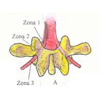
No que diz respeito a localização a estenose lombar pode ser central ou lateral. Na estenose lateral existe a diminuição do canal radicular que pode ser dividido em 3 zonas. A zona 1 é a área situada sob a apófise articular superior, medial ao pedículo, chamada de zona sub-articular; a zona 2 é a zona foraminal; a zona 3 é a zona extra foraminal.


O segmento anatômico pode ser dividido em 3 níveis:

1. Nível disco
2. Nível forame
3. Nível pedículo

As alterações que provocam a estenose do canal lombar ocorrem no nível do disco. Neste nível as estruturas que contribuem para a estenose do canal são: o ligamento amarelo, suas porções laminar e articular, as articulações interapofisárias e o disco intervertebral. Ao nível do disco, os fatores que são importantes para a compreensão da anatomia patológica lombar são: a forma do canal, onde há 3 formas, a de trevo, a triangular e a ovóide. Cerca de 15% da população tem a forma em trevo, que a forma que mais pré-dispõe as alterações degenerativas; alterações degenrativas como a protusão discal, hipertrofia ou pregueamento do ligamento amarelo e formações osteofitárias; e a espondilolistese que provoca um efeito guilhotina do canal vertebral. Todos fatores ocasionam a estenose do canal lombar, diminuindo o espaço útil para a cauda eqüina.


5.2. A HÉRNIA DISCAL

A hérnia de disco é uma das principais causas da Síndrome da cauda eqüina aguda. A hérnia ocorre devido a uma diminuição no conteúdo dos proteoglicanos no disco intervertebral, esses são os principais responsáveis pela hidratação do núcleo pulposo, dando a propriedade de gel do mesmo, fazendo com que as pressões no anel sejam distribuída de forma uniforme. Com diminuição da pressão de embebição do disco, maior pressão é transmitida as fibras do ânulo, o núcleo perde suas propriedades hidráulicas, de verdadeiro amortecedor das pressões, e as fibras do ânulo são mais susceptíveis a ruptura.

Qualquer fator como excesso de peso ou má postura leva a estrusão desse núcleo, dando origem a hérnia discal.

Hérnia de Disco


5.3. FRATURA
A fratura é considerada uma lesão traumática, esta pode ser de originada em acidentes automobilísticos, queda de altura, mergulhos, por ferimentos com armas brancas, com ferimento com arma de fogo (projétil).

Ao ocorrer um trauma vertebral, ou seja, uma fratura, uma ou mais vértebras podem se deslocar, provocando uma compressão sobre a cauda eqüina. Normalmente ocorre com as fraturas abaixo do nível de L2.


5.4. TUMOR E INFECÇÕES
Alguns tumores como ósseos, ou até mesmo tumor de vaso, podem comprimir a cauda eqüina causando lesão e comprometimento das funções dos órgãos e sistemas da pelve e movimentos e sensibilidade nos membros inferiores. Algumas infecções, causadas por vírus ou bactérias também podem acometer a cauda eqüina, desencadeando a Síndrome.


6. FISIOPATOLOGIA DA COMPRESSÃO DAS RAÍZES DA CAUDA EQÜINA
Foram feitos estudos para se descobrir as alterações provenientes da compressão na cauda eqüina. O experimento foi realizado em animais.

Observando um modelo experimental de compressão da cauda eqüina no porco, Olmarker e Cols. Mostraram alterações na circulação venosa com pressões de 10mmHg. Alterações da condução neural foram obtidas com pressões de 50-74mmHg após 2 horas de compressão. Com pressões de 100-200mmHg havia bloqueio total da condução neural, com grau variado de recuperação quando se retirava a compressão. As alterações da propagação do impulso nervoso são dependentes da pressão sanguínea, sendo que o limiar de propagação do impulso diminui com a hipotensão.

Freqüentemente a estenose central ocorre em mais de um nível, e existe a associação da estenose central com a lateral. Nestes casos pode haver um distúrbio maior da nutrição e função das raízes.


7. QUADRO CLÍNICO
O quadro clínico clássico é o da anestesia em sela, retenção urinária, obstipação, ciatalgia contínua e anestesia plantar. Nestes casos, existe anestesia na distribuição dos nervos sacrais (nádegas, períneo, escroto, lábios, pênis e clitóris) e dependendo do nível de compressão, também se pode observar diminuição da sensibilidade nas pernas e pés. O exame físico mostra ausência do reflexo anal e bulbocavernoso, com graus variados de hiporreflexia e de paresia dos membros inferiores. A paralisia da bexiga pode ocorrer sem dor no membro inferior e a ciatalgia pode ser unilateral. O sinal de Lasegue bilateral pode ser positivo. No exame do abdome pode haver distensão da bexiga.

Na Síndrome da Cauda Eqüina crônica os achados clínicos são mais sutis. Pode haver diminuição do tônus retal e a sensibilidade perineal estar alterada. Os achados do exame neurológico podem ser normais em repouso e presentes com o caminhar ou com a manobra de extensão da coluna lombar. Pode ocorrer associação de dor na virilha perineal, vulvar ou testicular, incontinência urinária ou urgência miccional de início gradual durando meses ou nos.

Esses sintomas podem não estar associados à claudicação neurogênica. É comum a lombociatalgia crônica.
A dor pode ser causada pela compressão aguda ou dilaceramento das raízes nervosas. Freqüentemente a dor de raiz nervosa é descrita como aguda, lancinante, em queimação, ou penetrante, e tipicamente segue um padrão de dermátomos, ocorre muito nesta Síndrome pela elevada distribuição de raízes nervosas.

O tratamento da dor de raiz nervosa é um problema clínico perturbador. Foram sugeridas abordagens múltiplas, com graus variáveis de êxito. O tratamento conservador envolve a terapia medicamentosa e a estimulação elétrica nervosa cutânea. Intervenções cirúrgicas para a dor mais grave e debilitante são: secções da raiz nervosa (neurectomias) e rizotomias posteriores.


8. DIAGNÓSTICO
A história e o exame físico fazem a suspeita diagnóstica da Síndrome da Cauda Eqüina. Existe uma série de exames subsidiários que nos permite confirmar o diagnóstico, bem como planejar o tratamento. Os exames são radiografia simples, a tomografia computadorizada axial, a ressonância magnética e os estudos eletrofisiológicos. No raio X, o médico poderá observar comprometimento das raízes nervosas, por alterações degenerativas, como formação osteofitárias, retrolistese, estenoses e outras.

A mielografia seguida de uma tomografia computadorizada axial tem sido o exame mais usado para o diagnóstico, quando se trata da Síndrome da Cauda Eqüina proveniente de uma estenose lombar. A tomografia computadorizada é um bom método para avaliação das causas ósseas e partes moles. Detecta alterações como o canal em trevo, hipertrofia das alterações interapofisárias, algumas deformidades como a escoliose que pode estar promovendo uma compressão das raízes da cauda, fragmentos de fratura e fraturas pós-truma. Em geral a Tomografia computadoriza é realizada entre os níveis de L3 - S1 em cortes axiais de 5 mm. Nos casos de estenose o rastreamento tem que ser realizado de L1 - S1.

A ressonância nuclear magnética é o único exame que permite a aquisição de imagens em vários planos, sendo os mais usados os planos sagitais e axiais.

A eletroneuromiografia é muito importante e útil, pois através desse exame podemos observar o grau de comprometimento da raiz nervosa.

No caso de possível hérnia discal, o diagnóstico é dado através da ressonância nuclear magnética ou a tomomielografia.


8.1 DETERMINAÇÃO DO NÍVEL NEUROLÓGICO
A American Spinal Injury Association (ASIA), desenvolveu um protocolo padronizado. Os pontos chaves são testados com um alfinete e com um chumaço de algodão.

* O alfinete - Determina o agudo e o rombo.
* O algodão - localizar o tato ligeiro.

São recomendados ainda testes de sensibilidade profunda e da posição. Os resultados dos testes são listados no protocolo.
Existe uma classificação chamada Medida de Independência Funcional (MIF) foi adicionada recentemente no processo de avaliação do paciente. Este método é usado para monitorizar e avaliar o progresso com tratamento. Ele mede as atividades de vida diária nas áreas de cuidados pessoais, controle esfincteriano, mobilidade, locomoção e comunicação social. Atividades como o comer, higiene pessoal e o vestir são calculadas numa escala que mede o nível de dependência / independência.

Realizar um exame neurológico acurado e completo e determinar o nível neurológico da lesão ajuda a estabelecer metas reais para um programa de reabilitação que vise proporcionar maior independência ao paciente e promover melhor reintegração do mesmo dentro de sua comunidade.


9. PROGNÓSTICO
O potencial para a recuperação da lesão esta diretamente relacionado a extensão da lesão. Nos casos de lesões incompletas, que é a mais comum na Síndrome, é observada alguma evidência de função sensitiva e/ou motora abaixo do nível da lesão medular, depois que o choque espinhal cedeu. Os sinais iniciais de uma lesão incompleta podem estar indicados por um "sacro preservado" (sensação perianal, tônus no esfíncter retal, ou flexão ativa dos dedos dos pés). As lesões incompletas também podem apresentar-se com pontos ou áreas disseminadas de função sensitiva e motora.

É importante observar que na maioria dos casos de lesões incompletas a melhora começa que imediatamente após a cessação do choque espinhal. Muitos pacientes terão alguma melhora progressiva do retorno muscular. Pode ser mínima, ou, menos freqüentemente, dramática, usualmente se tornando aparente durante os primeiros (e diversos) meses após a lesão. Com uma consistente progressão do retorno da função (diária, semanal ou mensalmente), maior recuperação poderá ser esperada ao nível desta mesma velocidade, ou numa base ligeiramente mais lenta. Avaliações meticulosas e freqüentes das funções sensitivas e motoras que neste período propiciarão importantes informações para o progresso da recuperação.

Com o tempo, a velocidade de recuperação cairá, sendo alcançado um platô. Quando este platô é atingido, e não é observada atividade muscular por diversas semanas ou meses, não deve esperar que no futuro ocorram recuperações adicionais.


10. TRATAMENTO
O tratamento dos pacientes com lesão na cauda eqüina necessita de consideração especial.


10.1 TRATAMENTO CONVENCIONAL
No tratamento convencional é realizada a manobra com a manipulação de Crede / Valsalva, para que os pacientes possam urinar, devido a presença de uma bexiga arreflexiva (não contrátil).

Existem pacientes que através de uma avaliação urodinâmica mostraram esfíncter uretral externo intacto, e, portanto esses pacientes podem conseguir urinar gerando altas pressões intravesicais.

A catetização intermitente é começada a cada quatro ou seis horas, e com restrição hídrica a cerca de 1,5 litro por dia, esses pacientes podem ser tratadas com três catetizações por dia.

A catetização intermitente pode ocasionar uma infecção. Neste caso é indicado o uso de antibiótico adequado principalmente nos primeiros 3 dias, a fim de prevenir a disseminação da infecção uretral.

A fim de reduzir a incontinência de esforço. A paciente é encorajada e treinada para esvaziar a bexiga pela manobra de Crede /
Valsalva periodicamente. O fracasso do tratamento com catetização intermitente usualmente resulta em colocação em longo prazo de um cateter de demora.

Procedimentos neurocirúrgicos tais como bloqueios seletivos de raízes nervosas sacras e rizotomias podem ser indicadas a fim de converter uma bexiga reflexa (contrátil) em bexiga arrefléxica, a qual, conforme mencionado acima, pode ser facilmente controlada pela manobra de Crede/Valsalva.

Pode ser administrado anti-inflamatórios e analgésicos. Os anti-inflamatórios são de origem não hormonal. Todos os anti-inflamatórios podem ter efeitos colaterais sendo os mais comuns os gastrointestinais e renais.

O uso de analgésico é importante tanto na fase aguda como crônica.

Deve ser feito a descompressão dos elementos neurais através da cirurgia. No caso de uma hérnia de disco unilateral é realizada uma laminectomia unilateral.

Quando existe uma estenose associada, a laminectomia é bilateral.

Nos casos agudos a cirurgia deve ser de urgência, e os melhores resultados cirúrgicos ocorrem nos casos operados nos três primeiros dias do início do quadro. Não houve correlação entre a duração do quadro clínico e o resultado cirúrgico. Nos casos de Síndrome da Cauda Eqüina crônica a descompressão não deve ser feita de urgência e o Paciente deve ter uma avaliação clínica completa. Nos casos agudos raras vezes o paciente se recupera totalmente, independente do intervalo entre o início do quadro e a descompressão cirúrgica. A ciatalgia bilateral é considerada sinal de mau prognóstico. A persistência da anestesia em sela é sinal que a função esfincteriana persistirá alterada. Nos casos crônicos a recuperação é lenta e incompleta. Deve-se explicar ao paciente que a cirurgia tem como objetivo impedir a piora do quadro clínico.


Fase Aguda
O esvaziamento intestinal também deve ser realizado através da estimulação proveniente de massagens abdominais no sentido horário ou uso de estimulação elétrica transcutânea.

Quando houver a volta da atividade intestinal deve-se introduzir o estímulo retal com supositórios neutros ou toque retal diariamente, após as grandes refeições. A dieta deve ser equilibrada é rica em fibras, acompanhada de uma ingestão líquida diária, em torno de 2 litros. Quando as manobras forem ineficazes, utiliza-se laxativos suaves por agentes ativos de superfície.


Fase Crônica
Iniciar precocemente a reeducação intestinal na posição sentada, que favorece o esvaziamento intestinal. Manter ingestão hídrica diária em torno de 2000 ml, de acordo com o programa de reeducação vesical e dieta hipercalórica, hiperprotéica e rica em fibras.
O uso de supositórios de glicerina e estimulação digital deve ser mantido nesta fase para se estimular os reflexos de evacuação.
Deve-se instituir horários regulares de refeições. No treinamento intestinal e regulação e regularidade dos movimentos intestinais são os objetivos mais importantes. Deve-se buscar a continência e a atividade intestinal socialmente aceitável e evacuações regulares com fezes de consistência adequada. O hábito intestinal pode ser diário, em dias alternados ou a cada três dias.

A manobra de Crede para os homens é recomendável que seja feita sentado para o esvaziamento vesical e o intestinal simultaneamente.

Esse programa deve ser mantido em períodos longos para se obter uma boa reeducação intestinal.

No que diz respeito ao tratamento para as disfunções sexuais é feito injeções de papaverina ou implantes cirúrgicos, no caso dos homens. Com o intuito de promover uma melhor ereção. Nas mulheres pode-se fazer o uso de lubrificantes artificiais. As alternativas para se manter um ato sexual, como beijos, sexo orogenital, uso de estímulos manuais em áreas genitais e não genitais vibradores podem e devem ser largamente estimulados e usados para que se tenha satisfação sexual em ambos os sexos.

Obs.: Na fase aguda o paciente deve fazer repouso de uma semana. Com a melhora do quadro e principalmente da dor orienta-se o paciente para controlar sua atividade física na medida de suas possibilidades.


10.1.1 Retreinamento Intestinal
Os pacientes que saíram da fase de choque, na Síndrome da Cauda Eqüina, tratam seu intestino com remoção manual em uma escala diária ou em dias alternados.

Esses pacientes podem começar a ter evacuações intestinais acidentais quando começam a usar um aparelho tutor ou a caminhar nas paralelas. Aumentar a freqüência dos cuidados intestinais de dias alternados para diariamente, acompanhando por instruções para manter fezes firmes através do controle da dieta, pode eliminar os acidentes.

Quando a tolerância a sentar-se permitir, o paciente deve receber tratamento intestinal em uma cadeira higiênica para permitir que a gravidade ajude na evacuação intestinal. A massagem do abdômen a partir da direita para a esquerda também pode facilitar a movimentação das fezes para o trato inferior. Nas ocasiões em que não são evacuadas fezes com os métodos convencionais, o paciente pode necessitar experimentar um laxante brando e repetir o programa intestinal 8 a 10 horas mais tarde.


10.1.2 Acompanhamento
Todos os pacientes necessitam seguimento urológico periódico pelo resto da vida. Durante o primeiro ano após a alta, revê-se o nitrogênio uréico sanguíneo, urucultura (e sensibilidades), e o bem estar geral do paciente. Catetizações desnecessárias para verificar a urina residual em alguém que não mostre nenhuma evidência clínica de retenção de urina, infecção ou litíase podem não ser justificadas. Em pacientes assintomáticos sob os demais aspectos cuja urodinâmica foi anteriormente estudada, em vez de pielografia intravenosa anual de rotina, a renografia por radioisótopos para função renal, sonografia e radiografia simples do abdômen podem ser suficientes para o acompanhamento. A sonografia transretal linear é considerada muito útil, como uma modalidade não invasiva, para o acompanhamento visando determinar a urina residual e qualquer obstrução vesical.


10.2 TRATAMENTO FISIOTERÁPICO
O tratamento mais utilizado é o conservador, que é um tratamento repetitivo e requer um alto grau de disciplina, perseverança e motivação, precisando muitas vezes da interferência do fisioterapeuta não só como especialista mais também como incentivador para que trabalho seja realizado com sucesso.

Os tratamentos conservadores consistem em exercícios e eletroestimulação. As contrações simples do assoalho pélvico podem ser ensinadas e praticadas, desde que o paciente seja capaz de contrair voluntariamente a musculatura pélvica.Com prevenção pode ser usado um perineômetro (dispositivo usado para medir a pressão dentro da vagina) para evitar a resistência e melhora e o biofeedback motiva a prática dos exercícios.


10.2.1 Exercícios de Contração do Assoalho Pélvico
Antes de instruir o paciente sobre o modo de realizar as contrações é preciso determinar, além de qualquer dúvida se o paciente é capaz ou não de ativar voluntariamente os músculos corretos. As contrações dos músculos glúteos, adutor do quadril e abdominal, a retenção fôlego e até o abanamento do abdome tem sido confundido com contrações da musculatura do assoalho pélvico. A capacidade de contrair os músculos elevadores do ânus deve tecnicamente ser estabelecido de maneira objetiva pelo fisioterapeuta, como parte do exame inicial.

Se após uma cuidadosa instrução houver incapacidade de contração voluntária, deve-se tomar providências para reeduca-la. Isso, usando os meios adicionais que mais se adequarem, por exemplo, estimulação elétrica.

O ensino das contrações do assoalho pélvico, para o paciente praticar de modo regular sem auxílio é uma tarefa muito difícil exigida pelo fisioterapeuta já que os músculos não estão diretamente visíveis.

Na posição inicial as contrações podem ser feitas em qualquer posição, porém, a mais adequada é estar sentada em uma cadeira dura e inclina-se para frente para apoiar os antebraços nos joelhos, com as coxas e pés afastados. O períneo fica contra o assento da cadeira, de modo que há um feedback do estímulo sensorial do períneo, e uma mudança de sensação é geralmente visível sobre a saída pélvica durante a contração.

Na primeira sessão pede-se ao paciente para manter uma contração com força até que ele sinta a fraqueza muscular.
A linguagem de estímulo deve ser escolhida de modo específico para cada paciente utilizando palavras e imagens de fácil entendimento

* Comprima, solta, comprima, solta;
* Comprima o assoalho para cima e para baixo, segure e solte;
* Interromper a urina;
* Tentar contrair toda vez que espirrar, tossir ou rir pode evitar vazamento, quando os músculos estiverem mais hipertônicos.

Se sentir dificuldade estando sentado ou deitado realizar exercícios em pé com pernas ligeiramente afastadas.

O paciente precisa de estímulos regulares para aumentar a extensão, intensidade e o número de contrações repetidas. Deve ser usado um certo número de posições, trabalhando mais as que costuma ocorrer vazamento.


10.2.1.1 Incontinência Fecal
Quando o paciente se queixa de incontinência amorretal ou de urgência para defecar é muito importante a intervenção de um tratamento adequado, por é um problema embaraçoso que leva o paciente a situações desagradáveis e até mesmo o afastamento do convívio social. Alguma recuperação pode ocorrer com exercícios regulares do assoalho pélvico a resistência do mesmo pode chegar a um nível razoável. Caso não haja capacidade de contração devem ser utilizados métodos interferenciais que são os mesmos utilizados na incontinência urinária, porém não apresentando tanto êxito. Já o estímulo com eletrodo anel tem mostrado ser benéfico (Scokier, 1989). Podendo ser um recurso útil no auxílio do tratamento tanto como dispositivo do biofeedback para ensinar o controle do esfíncter anel de início, como um método para fortalecer os músculos.

A contração do músculo elevador do ânus inibe diretamente o centro de micção e a urgência de esvaziar a bexiga pode ser controlada.


10.2.1.2 Biofeedback
É um equipamento que é conectado no paciente por sensores onde se solicita ao paciente a contração da musculatura do assoalho pélvico e os sinais de atividade elétrica dos músculos são mostradas em um monitor em proporção a intensidade e duração da contração dando ao terapeuta e ao paciente o feedback do comportamento da musculatura permitindo a reeducação e obtenção de resultados satisfatórios, qualidade da musculatura, aumento do controle miccional e fecal e aumento do prazer sexual. É esperado que eu seria para motivar o paciente à prática e também agir no sentido de fazer contrações mais fortes e mais longos.

É contra-indicado em hipertonia total do assoalho pélvico já que é necessário a contração da musculatura para que haja a transmissão da atividade elétrica para o monitor do aparelho.


10.2.1.3 Estímulo Muscular Elétrico
Em geral os fisioterapeutas têm utilizado a estimulação elétrica na reabilitação de pacientes que têm dificuldade em realizar a contração voluntária dos músculos do assoalho pélvico. Afirma-se que pela aplicação de sinais de corrente elétrica é possível reassumir o controle normal entre nervo e músculo em uma unidade motora.

A aplicação é feita por eletrodos nos métodos de quatro pólos e de dois pólos.


10.2.1.4 Tratamento Persistente
Quando apesar de todo tratamento repetido e exaustivo o paciente continua com algum grau de incontinência os esforços devem ser dirigidos no sentido do tratamento e achar a melhor solução para cada caso específico. O terapeuta pode ser capaz de produzir um pouco mais de resistência ou limite de movimento para permitir ao paciente tornar-se mais independente. Dando a ele condições de sair de casa e aproveitar a vida, pois a incontinência leva a uma privação social que desencadeia a depressão.


11. CONCLUSÃO
Neste estudo, concluímos que apesar da Síndrome da Cauda Equina ser uma patologia não muito conhecida até mesmo pelos elementos da área da saúde, tem-se ocorrido de certa forma freqüente. Esta ocorre de forma secundária a alterações, como as degenerativas da coluna vertebral, precisamente a nível lombo-sacral, fraturas, hérnias discais, tumores e outras.

A fisioterapia vem buscando meios de minimizar os efeitos decorrentes da Síndrome, bem como promover uma melhor qualidade de vida e o retorno precoce do paciente as suas atividades diárias.
Autores: Ana Beatriz P. do Santos, Cláudia Regina G. Ferreira, Daniel M. Rojas, Mônica P. Gomes, Silvania Oliveira

quarta-feira, 3 de agosto de 2011

Healthcare partnership pays big dividends

By working together, Blue Shield of California, Catholic Healthcare West and Hill Physicians Medical Group saved more than $20 million in costs last year in serving 41,500 HMO members in Northern California.By Duke Helfand, Los Angeles Times

A rare alliance of healthcare rivals — a giant insurance company, a major hospital chain and a large doctors group — has managed to reduce healthcare costs through a radical new strategy: working together.

The collaboration among Blue Shield of California, Catholic Healthcare West and Hill Physicians Medical Group shaved more than $20 million in costs last year and prevented an insurance rate hike for public sector workers in Northern California.
The three partners cite evidence that the quality of care also improved: Hospital stays were shorter, readmissions dropped and doctors and nurses kept closer tabs on patients.

Relationships between these kinds of companies are typically adversarial, with doctors and hospitals trying to negotiate higher prices for their services as insurers strive to limit what they pay out.

But driven by a mutual interest to cut costs and to be more competitive, the three devised a strategy they believed would deliver medical care more efficiently.

Skeptics worry that the partnership and others like it will put cost-cutting ahead of patient care. Healthcare experts believe, however, that such experiments — including one being planned in Orange County — hold important lessons for an expected wave of similar "accountable care organizations" as part of the nation's healthcare overhaul.

"The fact that they achieved substantial savings in the first year highlights the potential for the model," said Dr. Elliott Fisher of the Dartmouth Institute for Health Policy and Clinical Practice in New Hampshire. "There are a lot of opportunities to achieve savings, even in the short term."The three partners began planning their experiment in early 2008 — well before President Obama and Congress opened a heated national debate over how best to control healthcare spending.

Their talks centered on 41,500 members of a Blue Shield HMO who were served by Hill Physicians, whose doctors are affiliated with Catholic Healthcare West, the state's largest hospital chain.

All of the participants got their Blue Shield insurance through the California Public Employees' Retirement System and lived in Sacramento, El Dorado and Placer counties.

Senior executives from the three healthcare companies said they had to overcome past quarrels to collaborate.

"Our staffs had a history of combating with each other through negotiations," said John Wray, a senior vice president with Catholic Healthcare West. "We had to trust one another to make it happen. This was a very significant culture change between the organizations."

The partners overhauled procedures for medical treatment and hospital care. They started by taking the extraordinary step of sharing closely guarded financial and medical information.

They discovered, for example, that a handful of elective procedures — including weight-control measures — were among the biggest cost drivers.

They took action to cut these costs. Overweight patients, for instance, were given an opportunity to enroll in a Hill Physicians weight-loss program in which a psychotherapist and dietitian teach how to manage food cravings and make healthier eating choices.

The efforts helped reduce the surgeries by 13% last year, the hospital system reported.

Emergencies were another target. When patients were taken to hospitals outside of Catholic Healthcare West, they were stabilized and then directed back to the hospital system for lower-cost "in-network" treatment. In all, 113 patients in the experiment went to emergency rooms outside the network last year. Of them, 85 were transferred back to Catholic Healthcare West hospitals once stabilized.

Medical centers and doctors also took aim at expensive repeat visits for hospital patients by paying greater attention to follow-up care. As part of the hospitals' checkout procedures, nurses reviewed patients' post-hospital instructions and then asked them to repeat it all back. The providers also made sure patients had made appointments with doctors before going home.
As a result, hospital stays overall were shortened and the numbers of patients who had to be readmitted dropped by 15% in 2010, the providers said. Both results, they said, were signs of improved care: Spending less time in the hospital meant less chance of getting infections, while fewer readmissions meant patients remained healthier after surgery.

Of the $20 million in savings, Blue Shield recouped $15.5 million for a pledge it had made to CalPERS not to increase insurance rates for the 41,500 people in the experiment. The remaining $5 million was divvied up among the three partners. They also had agreed to share the risk if the experiment didn't pay off.

Blue Shield executives believe the rate cut helped attract about 1,000 new policyholders last year and gave the company a competitive edge as it vied for a new CalPERS contract. Blue Shield covers more than 400,000 CalPERS members, about one-third of those who get health insurance through the giant state pension fund.

"Clearly it was worth our while," said Juan Davila, Blue Shield's top executive who oversees provider contracting. "Our biggest client is happy with us."

Some healthcare advocates question whether patient health will suffer as Blue Shield and its partners look for additional cost savings. Could procedures or tests be denied because they are too expensive, they ask.

"You want to make sure that people have access to the care they need," said Anthony Wright, executive director of the consumer group Health Access California. "That's part of the balancing act."

The experiment's three partners insist that healthcare quality is as important as saving money.

CalPERS, meanwhile, said it is pleased to see improvements on both fronts, even though most of the savings in Northern California went unnoticed by individuals because the money was shared among the agency's entire statewide membership.

Blue Shield is joining with hospitals and medical groups for similar alliances in San Francisco, Modesto and Orange County. The insurer and St. Joseph Health System in Orange are gearing up to launch a collaboration in January for 30,000 Blue Shield HMO members. About one-quarter of them belong to CalPERS.

"We firmly think this is the right way to go," said Ann Boynton, a CalPERS benefits executive. The cost-cutting strategy, she said, "will boost care for our members and moderate the long-term increases in cost."

duke.helfand@latimes.com

quinta-feira, 28 de julho de 2011

B e t t e r P a t i e n t C a r e : V i r t u a l l y T h e r e

Autora: Judy Hanover  August 2010

This IDC Health Insights White Paper identifies the key benefits from desktop virtualization in the clinical environment and presents case studies from three hospitals that are early adopters of desktop virtualization in the clinical environment. The landscape for U.S. providers has been altered by two laws, the American Recovery and Reinvestment Act (ARRA) of 2009 and the Patient Protection and Affordable Care Act (PPACA) of 2010. These laws have created unprecedented incentives for the adoption of clinical technology and new goals for delivering higher-quality care to more Americans at lower cost. This will require providers to adopt new technologies rapidly and at the same time streamline their processes, cut costs, and improve quality. To do this, providers will be forced to examine all areas of their business and technology infrastructure. One of the key technologies that IDC Health Insights has identified for making provider IT departments more efficient and helping to meet these goals is virtualization. The return on investment (ROI) for provider organizations has been well documented for server and desktop  virtualization, but the clinical benefits that arise from the virtualization  of the clinical desktop have only recently begun to be discussed.
The research for this white paper included detailed interviews with IT  departments at three hospitals that are early adopters of desktop  virtualization for clinical applications. The observed clinical benefits  from desktop virtualization in these organizations include:

● Improving efficiency in the IT department, enabling the delivery of higher service levels, better uptime, and higher availability and performance of clinical applications while maintaining staff levels

● Driving adoption of clinical applications by improving the usability and performance of legacy clinical applications

● Supporting provider mobility with wireless access, thin-client hardware options, single sign-on, and session virtualization

In the rapidly changing environment created by the new incentives and regulations, IT efficiency and agility are critical to success for provider


I N   T H I S   W H I T E   P A P E R

This White Paper is presented by IDC Health Insights and sponsored by VMware. The objectives were to gain insights into:

● The decision-making process for investment in desktop virtualization of clinical application by hospitals

● The IT and clinical benefits experienced by three provider organizations that invested in VMware desktop virtualization solutions

● The importance of desktop virtualization in unleashing the value of electronic medical record (EMR) and other clinical applications

To meet these objectives, IDC Health Insights conducted three indepth interviews with provider organizations that have adopted desktop virtualization of clinical applications in the 2009–2010 time frame; the interviews were conducted in March–June 2010.

S I T U A T I O N   O V E R V I  E W

As the healthcare industry seeks to implement and drive widespread adoption of clinical computing, performance and accessibility of applications for clinicians are critical. In the hospital environment, solutions like desktop virtualization are increasingly playing a role in bridging the gap between available resources and satisfaction of enduser requirements.
Incentives created by ARRA and the additional goals of PPACA are resulting in a surge of new EMR and computerized physician order entry (CPOE) system implementations, alongside expanded adoption of existing clinical information systems, analytics, and revenue cycle solutions. The growth in adoption of clinical information systems will result in a proliferation of point-of-care computing, dramatically increasing the number of client machines that need to be supported by  IT organizations.
Client or desktop virtualization provides a tool that can help provider IT organizations to:

● Deliver enhanced performance, uptime, and availability to providers using clinical applications

● Drive efficiencies and improve service provided by IT staff
implementing, managing, and maintaining clinical systems
● Improve accessibility and productivity for mobile users of clinical
applications in complex care settings
● Create a foundation of a private cloud internally, virtualizing
servers, applications, networking, desktops, and applications
● Manage healthcare information technology (HIT) efficiently by
centralizing services in a single datacenter, thereby freeing up
resources that were devoted to catering to individual endpoint
devices
● Increase security by controlling information and keeping data off
endpoint devices
● Lower operating expenses by redirecting IT resources from help
desk calls to support new projects, EMR, and additional desktops for
clinical applications — all without increasing staff
S O L  U T I O N   D E S C R I  P T I O N
Current commercially available software products encompass three
types of virtualization: server virtualization, application virtualization,
and, most recently, client or desktop virtualization. This white paper
focuses on the features and benefits associated with virtualization of
the client.
D  e s  k t  o p   a n  d   A  p p l  i  c a t  i  o n   V  i  r  t  u a l  i  z  a t  i  o n
Client or desktop virtualization is a datacenter-centric computing
model that borrows from the traditional thin-client model but is
designed to give system administrators and end users the best of both
worlds: enabling system administrators to host and centrally manage
virtual and/or physical desktop machines in the datacenter while
giving end users the traditional PC desktop experience to which they
have become accustomed.
In virtualized client environments, the software image of a physical
desktop PC is replaced by a virtual PC running on a server. Each user
accesses a unique virtual PC, complete with its own virtual CPU,
RAM, and hard disk. Virtual PCs run concurrently on top of the
virtualization layer provided by the hypervisor software, which also
controls the computing resources (mainly CPU and RAM) allocated to
the virtual PCs. Although multiple virtual PCs typically run on a single
server, the crash of a single virtual PC is unlikely to cause other virtual
PCs on the same server to crash. Users access datacenter resources via
thin-client or full-client workstations.

Application virtualization separates applications from the underlying
operating system and packages them with a virtual operating system in
a completely isolated container. This allows users to run applications
on the same desktop that may have conflicted before or run
applications that use different Windows operating systems side by side
on a single desktop. Applications can be virtualized and delivered into
virtual desktops or traditional desktops.
V M w a r e   S o l u t i o  n   D e s c r i p t i o n
VMware provides a number of solutions to accomplish virtualization
implementations at the hospitals we spoke with. The VMware products
in use include:
● VMware View provides a centralized virtual desktop and
distributed virtual desktop that allow remote and local management
of fully virtualized desktops.
● VMware ThinApp is a key component of VMware View and is also
available as a standalone solution. This solution provides application
virtualization to simplify application delivery and management.
The three hospitals discussed in this study are using VMware View
and ThinApp to deliver client virtualization solutions.
B E N E F I  T S
Enterprises have discovered that the use of virtualization to support
desktop workloads, like the use of virtualization in support of
traditional server workloads, creates a range of significant benefits.
These benefits include improved IT management efficiency, improved
price efficiencies, and improved functional capabilities. The key IT
benefits associated with desktop virtualization in the healthcare
industry fall into the following areas:
● Enablement of thin clients. Because little computational execution
occurs at the edge in a desktop virtualization environment, the
computing architecture becomes less reliant upon the horsepower in
endpoint devices. This creates an opportunity for IT to significantly
drive down the cost of endpoint hardware either by extending the
life span of existing PCs by repurposing them as client endpoints or
by replacing aging PCs with a thin-client device, which typically
operates across a life span twice that of a standard PC.
● Improved data security. The ability to move data from the edge
of the IT environment into the datacenter inherently reduces the
security risks to an IT organization. Centralization of data access
can mitigate the risk of data leakage and theft and simplify
compliance procedures.

● Simplified data backup. Because centralized virtual desktops
reside entirely within the datacenter, it is easier to ensure full
compliance with backup policies. Furthermore, depending on how
the platform is architected, the use of consolidated images and
delta files may further simplify the abstraction and collection of
important data, thereby simplifying backup processes.
● Simplified disaster recovery.  Virtual machines (VMs)
significantly simplify disaster recovery because central IT staff can
easily revert virtual desktops back to their last known good states.
Thus, IT no longer needs to provide spare endpoints that are up to
date with the latest image.
● Time to deployment.  In particular, when thin clients are used
within a virtualized architecture, the process around deployment is
significantly simplified because nothing is installed on the
endpoint device.
● Simplified PC maintenance.  When used appropriately, virtual
desktops can be far easier to maintain than traditional PCs. Because
of the unique characteristics of virtual machines, it can become quite
simple to patch applications, provision/deprovision users, migrate to
new operating systems, and perform auditing duties.
● Flexibility of access. Because corporate desktop environments are
centralized, access to them can be provided to users who do not
have access to their own PCs, in situations where users need secure
access to clinical systems while working from home, on hospital
floors, in acute care environments, or in other remote situations.
This is particularly important for mobile caregivers who work in or
move between hospitals, ambulatory care environments, and other
settings.
● Simplified application delivery and management. Applications
virtualized into single image executables can be centrally managed
and delivered to many endpoint devices. Application virtualization
also minimizes application testing and help desk support because
the application is completely isolated from the operating system,
ensuring the same application behavior independent of the
operating system configurations.
While IT improvements are certainly impressive in their own right,
enhancing the quality of care and patient safety is a top priority for the
healthcare industry. Desktop virtualization can be a critical component
of strategies to achieve these goals and deliver clinical benefits in
healthcare. The clinical benefits seen by the three organizations we
spoke with are discussed in the case studies presented in the following
section.

D  e s  k t  o p   V  i  r  t  u a l  i  z  a t  i  o n   a t   T h r  e  e   H e a l  t  h c  a r  e  O r  g a n i  z  a t  i  o n s  Case Study #1: University of Toledo

The University of Toledo (UT) went through a profound change four years ago when a 20,000-student university merged with a 300-bed teaching hospital. The opportunities presented by such a merger seemed endless. But with those opportunities came challenges; merging cultures, departments, and of course, technology was no easy task. There are more than 10,000 Windows desktops in the combined institution and over a dozen Information Technology support groups managing them. Josh Spencer, Team Lead for the Desktop Development group in Information Technology, provides desktop administration support such as application packaging, imaging, and desktop automation for more than half of these PCs. He identified a common challenge that plagues each of these groups: "maintaining stable desktop images and keeping them updated with the ever-changing software suites." A variety of methods have traditionally been used at UT, ranging from group policies to scripting upgrade routines to "sneakernet." According to Spencer, "This is particularly challenging because unlike some industries which require a handful of applications to do business, the IT groups at UT have to support hundreds of software titles." Any number and combination of applications might be required in student labs, in clinics, and in training rooms. The sheer number of applications and computers makes it difficult to keep the images current. The endless varieties of software combinations introduce instability. Spencer's team has implemented VMware View and ThinApp in both clinical and academic environments to address these challenges.


B e n  e f  i t s   o f   A p  p l  i c  a t  i  o n   V i  r t  u a  l i z  a t  i  o  n

 Over the years, the Desktop Development group has implemented a variety of application packaging technologies in an attempt to reduce the age-old problem of dll and registry conflicts. While incremental improvements have been realized, the group has found that these technologies all fall short, causing a great deal of administrative overhead to manage application conflicts and a loss of productivity for end users.  About three years ago, the group decided to try application virtualization using VMware ThinApp. This technology has two key components that made it a success: application isolation, which improved desktop stability, and application streaming, which reduced administrative overhead and increased uptime. According to Spencer, "Application isolation not only allows us to segregate the application and all of its dlls; we can actually include all supporting applications in the virtual 'bubble.' This concept introduces a level of stability never before attainable in our desktop environment."  Today, the Desktop Development group has approximately40 virtualized applications in production, the majority of which are in the clinical environment. The clinical staff at the UT Medical Center use applications such as McKesson Star Navigator and Care Manager and Allscripts Vision. These applications have all been virtualized and are streamed to thousands of physical and virtual desktops throughout the institution. "We started with our most complex and problematic applications and saw the benefits of application virtualization immediately," said Spencer. Applications such as Allscripts Vision require a specific set of prerequisite software such as Java, .NET framework, and IBM Informix Connect. Managing each of these applications independently and ensuring that they remained at the appropriate level to support Allscripts Vision proved to be a challenge. Spencer noted, "We simply put all the prerequisite software in the bubble with [Allscripts] Vision, and we don't have to worry about what is or isn't on the host; the application just works." Another improvement in stability comes from the fact that once virtualized, applications that would normally require local administrative privileges to function can run as a standard user. Spencer's team is now deploying clinical desktops with restricted privileges, preventing unauthorized changes and improving uptime.
Administrative overhead for deploying and upgrading applications has been reduced dramatically through the use of virtualized applications.
Rather than using traditional install/upgrade methods, Spencer's team is streaming applications from a central server. This method reduces the need to make changes on the endpoint computers. Spencer said, "We no longer have to write a scripted upgrade routine for our applications. We simply update an executable on the central share and the work is done.
We've seen our failure rates for an application upgrade drop from 5% to nothing; people sometimes don't even know we've done anything."
Clinical applications that have been altered by application packaging of any kind are usually not supported. Spencer's team made the decision that the benefits from repackaging outweighed the risk of diminished support from vendors and uses a traditionally installed version of the application when contacting suport.


B e n  e f  i t s   o f   D  e  s k t  o p   V i  r t  u a  l i z  a t  i  o n  

The University of Toledo used VMware View to build a private cloud infrastructure, providing students with on-campus and off-campus access to hundreds of virtual machines in dozens of unique configurations. Last year the university decided to implement 300 new virtual machines along with thin-client devices as part of an electronic medical record initiative in the Medical Center hospital. A variety of factors were involved in the decision, and several technological solutions were considered. In the end,Prior to the implementation there were approximately 600 PCs in the UT Medical Center hospital. The Horizon Electronic Documentation project required an additional 300 PCs to provide bedside patient care.
The work of deploying and managing these devices had to be done with no additional staff. Spencer's team worked with Clinical Informatics, Hospital Client Services, Server Administration, Network Engineering, and other IT groups to design a system that met the needs of the clinicians and that could be supported with existing human resources.
Wyse thin-client devices with custom XP embedded images were deployed to wall-mounted arms in patient rooms, desktops in nursing units, and mobile carts. Barcode scanners were attached to most devices for use in bedside drug administration. The thin-client devices were programmed to connect only to the VMware View servers.
The benefits realized from virtualizing the desktop environment at the University of Toledo Medical Center include:

● Removal of data from the endpoint device. Traditional desktop PCs allow end users to store sensitive information on the local drive, creating a potential risk in the case of theft. The thinclient/View model keeps all data in the datacenter and prevents anything from being stored locally.

● Reduced administrative overhead. Because applications on the virtual machines have been virtualized, allowing for more restrictive local privileges, desktop stability is greatly improved.

On the occasion that a VM does need to be reimaged, the use of VMware snapshots has reduced what takes more than an hour with a traditional PC to less than five minutes. "Our technicians can simply use the Virtual Center console to revert a VM to the snapshot taken when it was created. In a matter of minutes, the clinician is back up and running," said Spencer.

● Lower-cost endpoint devices. Thin clients are a fraction of the cost of a typical PC and have a longer life expectancy.

● Energy savings. Spencer reported that an internal power study shows a considerable reduction in energy consumption for thin clients and supporting back-end infrastructure compared with traditional PCs currently used in the hospital.

For the IT team at the University of Toledo, key learnings have included understanding how clinicians work, how they want to work, and what they need to be successful and then balancing all of that information with HIPAA requirements and the internal resources available to provide support.

Case Study #2: Overlake Hospital Medical Center Overlake Hospital Medical Center is a nonprofit community medical center that includes a 337-bed hospital and about 10 local clinics.


Overlake uses MEDITECH for its inpatient clinical and administrative systems and plans to upgrade to MEDITECH Client/Server v6.0 in August 2010. The outpatient clinics run eClinicalWorks for practice management and EMR, and in addition to the 10 hospital-owned clinics, an additional 1,000 community physicians are supported for a total of roughly 3,000 end users. Director of Technology and Information Security Officer Joseph Wolfgram will deploy desktop virtualization to 900 desktops out of his 1,800 workstations in 2010 and expects to see substantial future growth and the need to add additional endpoint hardware to support EMR users including clinical documentation.
Before selecting VMware, Wolfgram conducted a technology evaluation and vendor selection process, as well as a formal proof-ofconcept process, and looked at multiple vendors. Overlake's criteria for selection included the ability to deliver a virtual desktop with a user experience identical to that of a local workstation, with no delays or pixelation, and the ability to offer full-screen, slow-motion video without pixelation or chop. After a successful proof of concept using PC over IP display technology on a virtualized client, he began a VMware standard evaluation, assessment, and deployment project. Today, about 100 users at Overlake use a VM every day, the new clinical system is being built on the VMs, and Wolfgram is deploying virtualized production servers and storage. All EMR training and eventually clinical documentation at bedside will use the virtual machines.

C l i n i c a l   a n d   A d  m i n  i s t  r a t i v e   B e n  e f  i t s

Overlake plans to use VMware View to deploy MEDITECH systems for EMR and CPOE, as well as the eClinicalWorks practice management and EMR in its affiliated clinics. While the clinics currently use tablet PCs as their thin client, the virtualized system may deploy on the iPad in the future, which Wolfgram expects to be more reliable than the PCs currently in use. According to Wolfgram, "Any clinical application that we can move to a virtualized desktop, the plan is to move it there. I'm not aware of any specific technical limitations today that we've run across for any of our 160 applications we support." Like his peers at the University of Toledo, Wolfgram has run into licensing and support limitations. Wolfgram finds that license offerings from vendors lack options that are efficient for the virtualized environment, and he prefers to duplicate problems on a nonvirtualized machine to ensure they are not related to the environment before accessing vendor support. He uses NetApp storage and a "snapshotting" feature to move the applications from a virtual  machine to a physical machine quickly.


The benefits that clinicians at Overlake have seen from desktop virtualization include:

● User experience on virtual machine that is identical to user experience on local machine. In order for desktop virtualization to be successful, the user experience with the virtual machine needed to be identical to the user experience on a local machine. For example, start menus and scrolling on the virtual machine needed to behave identically to those on the traditional, local desktop.

● Video capabilities for training and patient education. Fullmotion video is delivered to the virtual machines without chop or pixelation, allowing clinical staff to complete annual training requirements that are delivered as video segments and to make videos available to patients. Although Overlake is not using telemedicine, videoconferencing tools such as Skype are being used to connect remote clinics and deliver information via video.

● Reduced cost. Overlake will realize cost savings by using virtualized servers and storage, in addition to using desktop virtualization to lower costs associated with thin-client workstations.

● Faster log-ins and mobility for clinicians. The upgraded clinical systems will provide advanced functionality and improve security, but at the same time they will require individual log-ins to replace shared workstations, which was expected to add to log-in delays for both Windows and the clinical systems. Mobile clinicians in Overlake's hospitals may need to log in hundreds of times during a shift, making these delays quite significant. The virtualized desktops will follow the clinicians, allowing for quick connections and disconnections, while the desktops and functionality follow the provider.

● Easier desktop management and faster software delivery. From the IT perspective, it will be easier to manage desktops, and staff will be able to deliver and maintain software more efficiently.

● Improved security. With a virtualized desktop, the data never leaves the datacenter, and patient data and sensitive information is not resident on the end devices. This will replace a slew of point solutions that are currently in place and maintained on local machines, such as local firewalls and encryption, creating a win for maintenance, support, and security.

B e n  e f  i t s   f  r o m   D  e  s k  t o  p   V i  r t  u  a l  i z  a t i o  n

At Overlake, IT goals include virtualization of clinical workstations  as well as administrative workstations. The number of virtualized clinical workstations is expected to reach 900 initially, but it could grow to 1,100 with the implementation of the new EMR and could reach  2,000 across 12 facilities with the added administrative workstations.

Phase 1 of the effort will virtualize about 80 clinical and business applications that are delivered on individual user virtual desktops.  Overlake's clinical IT environment includes about 90 applications that will be virtualized. Applications that will be virtualized later include financial systems and departmental systems for emergency and surgery. Some legacy clinical applications will continue to run outside the virtualized environment as Overlake considers the value proposition for virtualization. According to Wolfgram, a few clinical applications have "really strict access control criteria and run on proprietary hardware, so many of our clinical systems that are like that, we have no plans to virtualize it, even if we could technically make it work, it doesn't make sense, too much effort."
Physician buy-in has been a focus at Overlake. According to Wolfgram, "For the EMR, the MEDITECH upgrade, we've got a physician champion, and we are working very closely with him to make sure he's exposed to our piloting. He was in on the proof of concept. He's giving us guidance in terms of what our physicians are expecting to see out on the floor — are the connects and disconnects fast enough, is it running across enough platforms including mobile devices like the iPad, that sort of thing. So we're in the throes right now of gaining buy-in from the physician community." Physicians' concerns about the workflow changes from the EMR clearly outweigh those about the virtualized environment. "They actually see the virtualized desktop as something that's going to be saving a lot of pain for them because without it, there would be a lot of really slow log-ins that would need to take place," said Wolfgram.

One of the lessons learned about virtualization at Overlake included understanding the costs. In the virtualized environment, it was important to have high-speed servers, a high-bandwidth network, and significant available storage, which added to the costs of hardware — mainly servers, disks, and networking equipment — to support the virtualized environment and achieve the required performance levels.  A local implementation partner at Overlake also played an important role. Wolfgram emphasized that "it's critical to have an implementation partner that has done virtualized desktop deployments in production and in a healthcare setting. There are things that are unique to this environment, and you want an organization that has had experience, not just the education of virtualized desktop implementation, because there are a lot of technical details that don't exist in the traditional desktop environment that really matter in a virtualized environment … having an implementation partner that has kind of blazed their trail before you that's helping you avoid potential problems, I think that's critical."

Clinical benefits from virtualization at Overlake have included:

● Faster log-in times for providers. The ability to log in quickly and easily improves the productivity of providers who must log in from different locations many times during the workday. ● Continuous sessions. Provider sessions are continuous when the
providers change locations because they can reconnect to an
existing virtual machine in a new location and pick up where they
left off, improving productivity.
● Remote access. Compared with existing systems, the virtualized
desktop provides a significant improvement to the performance of
the clinical applications when accessed remotely. Providers may
access their desktops from any location with the Internet and a
Web browser, and the access is faster and less complicated.
● Improved security. Security on the thin-client devices used in the
hospitals as well as for remote access has improved as a result of
centralizing data and information.
● Ease of use. Accessing systems is less complicated, response times
are faster, and thin-client devices are allowing providers to access
clinical data in a more mobile fashion.
Case Study #3: Phoebe Putney Memorial Hospital
The IT department at Phoebe Putney Memorial Hospital is responsible
for systems at 3 hospitals and about 15 clinic sites throughout southwest
Georgia. The three hospitals include Phoebe Main, a 450-bed acute care
hospital; the newly acquired Phoebe Sumter, with 74 beds; and
Phoebe Worth, a 25-bed critical access hospital. In addition, the IT team
is adding responsibility for hosting a MEDITECH system at the
Southwest Georgia Regional Medical Center in Cuthbert, Georgia.
The hospitals have different administrative and clinical systems, and
given the varying sizes of the hospitals, they have different goals
and requirements for their systems that prevent the standardization of
clinical and administrative systems.
Phoebe Main has the McKesson Horizon Inpatient Suite and Horizon
Ambulatory Care installed at its clinics. At Phoebe Main, CPOE
implementation is two-thirds complete, putting the hospital well on the
path to achieving meaningful use and qualifying for ARRA incentives.
The Phoebe Sumter and Phoebe Worth hospitals will be migrated from
Healthland to MEDITECH Client/Server v5.6, and the newly acquired
Southwest Georgia Regional Medical Center will also be migrating to
the upgraded version of MEDITECH, which will allow it to access
clinical applications including CPOE. All of the IT systems for the
hospitals will eventually be centralized at Phoebe Main, with
applications distributed to individual sites using server virtualization
and VMware View for desktop virtualization. Virtualizing the servers
has allowed the IT team to put 28 servers on 6 physical boxes, saving
considerable space and money.
The three rural MEDITECH hospitals will have a total of 800–900
users; about 100 people are using VMware View right now.
Each patient room at Phoebe Sumter will have a thin client using VMware View, and this hospital could eventually have 200–300
VMware View seats. At Phoebe Main, 5–6 of the McKesson
application servers were virtualized when the hospital upgraded to
version 10.1. In addition, home-based transcriptionists and coders and
the oncology department are using virtual desktops. A rollout planned
for this fall will include Horizon Expert Documentation as well as
wired Wyse thin clients in hospitals for nursing documentation.
Horizon Expert Orders physician documentation is planned for
rollout next year.
C l i n i c a l   a n d   A d  m i n  i s t  r a t i v e   B e n  e f  i t s
About two years ago, the IT team at Phoebe Putney first looked into
virtualization for its home transcriptionists when it needed to replace
an aging Citrix implementation. The team looked at multiple options
and considered the costs of training and hardware, alongside ease of
use, installation, and support. It determined that centralizing support
with the VMware solution offered the best option to support the
current needs of the remote users and the future needs for clinical
applications and growth in the hospitals. Additionally, a resourceintensive oncology application it was running in many remote clinics
did not support Citrix but could be optimized on VMware View. The
requirement to support the oncology application across a limitedbandwidth link was a key factor in the decision to use View.
The Phoebe Main hospital will eventually have 400–450 thin clients in
patient rooms to support Horizon Expert Documentation. The IT team
expects it to be easier to have the solid state thin client in the rooms
and run the operating system on the back end using View. Benefits to
nurses from using virtualized clients are expected to include:
● Reduced downtime. The thin clients will replace PCs and be
locked down and less likely to break or malfunction. Patches can
be done on the back end to the operating system, and users don't
need to worry about rebooting when patches are installed.
● Faster log-ins. Nurses will see improved speed of access and be
more efficient due to the addition of single sign-on and multiaccess
log-in.
● Improved mobility.  Users will have the ability to pick up open
sessions when they change locations, making them more efficient.
● Expanded functionality. Nursing documentation functionality has
been implemented at the bedside using the virtualized thin clients.
The hospital also has a medication administration system installed,
and nurses are able to scan medication barcodes and document
administration of medication using the thin clients. In the future, the
thin clients will also be used to implement CPOE for physicians.


B e n  e f  i t s   f  r o m   D  e  s k  t o  p   V i  r t  u  a l  i z  a t i o  n
Clinical testing has been done within departments and clinical
informatics using the thin clients and nursing documentation
applications. Although the system has not been rolled out on floors yet,
according to Michael Elder, Director of Technical Services, "…clinical
informatics has given us thumbs up on all the applications, the speed,
the efficiency of the unit … the application is working well, printing is
working, everything seems to be a go for that. So far the applications are
working well; the efficiency of it, speed, ease of use — all that's really
good. Integrating that single sign-on solution with it too is a big part of
that and all that seems to be working without any problems."
The IT team expects the providers to appreciate the speed, response
time, and mobility advantages of the virtualized solution. In addition,
the IT team expects to virtualize the physician portal that doctors use to
access PACS images and EMR data remotely, which will eliminate the
need to send technicians to provider offices to perform updates to the
software, which is currently installed locally at provider offices. To date,
the system has made early users, the home-based coders and oncology
clinics, more efficient. Home-based coders and transcriptionists report
that even when working from a setting without high-speed Internet
access, they have been able to work efficiently and experience less
downtime using View.
For the IT team at Phoebe Putney, key learnings have included the
need to educate users thoroughly about the process for using the
virtual desktop sessions in order to keep things running smoothly and
to test systems thoroughly before going live. The IT team also reports
significant ROI from the use of virtualization. In addition, the hospital
is reaching out to other hospitals in the region that are not part of the
health system to see if they want to partner to share resources and
access the hosted MEDITECH implementation.
F  U  T U R  E   O U T L O O K
IDC Health Insights expects to see extensive adoption of all types of
virtualization, including client virtualization and application
virtualization over the next three to five years. Growth in use of
virtualization will occur as healthcare organizations increasingly see
the benefits of virtualization and its ability to help them meet the
demands for efficiency and productivity that are resulting from new
healthcare reform and IT initiatives. Providers will begin to observe
their peers putting in place virtualization implementations; conducting
successful proof-of-concept projects; and putting virtualization into
production with new EMR/EHR, ordering, and other clinical systems.
IDC Health Insights also anticipates that the use of virtualization as an
infrastructure used to manage desktop environments will continue to expand with the growth in the capabilities and maturity of virtual
desktop platforms, alongside the compelling clinical benefits and ROI.
Early adopters of virtualization technology in healthcare will continue
to provide a proving ground for horizontal vendors that are moving
into the high-growth healthcare market. An increasing number of
centralized virtual desktop (CVD) platforms will become available to
healthcare organizations, driving competition in pricing as well as
products and features, including industry-specific functionality. The
willingness of legacy application vendors to support healthcare
information and clinical systems in a virtualized environment may
limit growth initially. However, it is expected that clinical vendors will
improve support for virtualization as the demand from providers grows
and as the ability of virtualization to provide performance, application
and data security, and complementary features that are not accessible
or difficult to improve in their legacy applications becomes clear.
Additionally, inpatient and ambulatory healthcare IT vendors will
begin to facilitate virtualized implementation, creating more seamless,
simple, and comprehensive platforms.
E S S E N T I A L   G U I D A N C E
Desktop or application virtualization may not be right for all
organizations or for some user populations in healthcare. Virtual
desktop infrastructure does add a layer of complexity to the existing
infrastructure that might deter some organizations from going ahead in a
short time frame. Clinical application vendors have hindered the
advance of virtualization across the industry — as many have been slow
to embrace virtualization or to support and/or certify the performance of
their products in a virtualized environment. However, this is more of an
issue with server virtualization than with desktop virtualization, and IT
executives in provider organizations have responded accordingly.
Care should be taken when implementing desktop virtualization to
ensure that it is an efficient approach for utilizing all of the application
services that a specific user, or class of users, is accustomed to
accessing. Applications that are graphic intensive, for example, may
not perform as well in a virtualized desktop environment.

While server virtualization may require a larger environment and
number of servers to reap benefits, desktop virtualization is not subject
to the same constraints. Application virtualization and repackaging may
complicate accessing vendor support for healthcare applications, but the
benefits may outweigh this risk for many healthcare organizations and
applications; as with any new technology implementation, an
assessment of the benefits and risks for the organization and its IT
infrastructure should be considered. The majority of provider
organizations support desktop environments sufficient to justify the
investment and see considerable economic and operational benefits.


C  O N C L  U S I O N
With stimulus funding in the mix, the outlook for investment in
clinical applications is strong. Although hospitals have been slow to
adopt EMR and CPOE technology and current adoption of
comprehensive EMR and CPOE is estimated to be below 20%, various
industry estimates predict that 50–60% of all U.S. providers will take
advantage of stimulus funding to install EMR and CPOE by 2016.
This will bring EMRs into use by the majority of providers and
dramatically increase the responsibility of hospital IT departments for
supporting clinical desktops and applications.
Tools like desktop virtualization and application virtualization will be
required in order to support these additional desktops while
maximizing the productivity of both IT and clinical resources. The
benefits that accrue from desktop virtualization projects include cost
savings that will help stretch IT resources and capabilities,
productivity enhancements for providers that will help ease the change
required to move to electronic clinical processes while keeping
providers mobile, and security measures that will help control data
access in the expanding clinical computing environment.