REHABILITATION EVALUATION AND TREATMENT IN PATIENTS WITH LOW BACK PAIN
Michael Kaplan, MD
ASSESSMENT OF LOW BACK PAIN SPINAL MOTION
• Accurate measurement is very important.
• Limitation of spinal motion correlates with the presence of lower back disability.
• Identification of palpable spasms and understanding nerve innervation are essential.1
PALPATION
• A positive Larson test, performed with the patient in the prone position, can indicate segmental instability common in degenerative disease of the lower lumbar discs.
• Active splinting of the segment reduces or eliminates the tenderness elicited with pressure over the spinous
processes, which is suggestive of segmental instability.
• Tenderness from soft tissue injuries persists despite active splinting.
• Muscle spasm is defined by the presence of a persistent, palpable increase in muscle tone accompanied by
localized tenderness.
• A digital rectal evaluation can detect pelvic floor myalgia or another pelvic pathology.
• Gentle and systematic palpation of the coccyx, sacrum, levator, ani, coccygeus, and piriformis muscles and their associated ligaments and attachments should be performed.
NEUROLOGIC ASSESSMENT
• Straight leg raising (SLR) tests should be performed to detect nerve root irritation. The classic positive SLR
test is a reproduction of radicular pain at 30°–40°.
• Radicular pain reproduced at greater angles represents less significant nerve root irritation.
• Back and leg pain can be produced in the absence of nerve root irritation.
• Nonradicular pain may be caused by soft tissue tightness or spasms in the back, glutei, or hamstrings.
• Even with a soft tissue pain source, the SLR can still be used as an index of improvement during treatment.
• A positive crossed SLR test has the highest correlation with myelographic findings of a herniated disc.
• Significant inconsistency observed during sitting and supine SLR tests may provide insight into the psychogenic processes.
• Electromyography is a valuable adjunct in delineation and confirmation of neurologic findings.2
SPECIAL TESTS
• The Hoover test is of special interest and suggests the detection of malingering because it indicates the recognition of submaximal effort. The jolt test is a provocative method used to document pain enhancement or radiation due to sudden mechanical loading of the erect spine. While standing on tiptoes, the patient is asked to suddenly drop to a flat foot position.
A positive jolt test is characterized by an exacerbation or radiation of pain.
• Leg length can be measured from the anterior superior iliac spine to the prominence of the medial malleoli (true leg length) or from the umbilicus to the medial malleoli (apparent leg length).
TRUNK STRENGTH
• Abdominal oblique muscles can be graded with the trunk rotated, as when a situp is performed.
• A similar method can be used to grade back extensors: lying prone with a pillow under the abdomen and hips, the patient extends the trunk and holds against resistance applied by the examiner.
RADIOLOGIC TESTS
• Plain radiography remains the cornerstone of radiologic tests.
• Plain radiography allows visualization of degenerative disc disease, spondylitis, compression fractures, metabolic bone disorders, bone tumors, congenital anomalies, and transitional vertebrae.
• Oblique views of the lumbosacral level can be added to visualize the facet and sacroiliac joints.
• Flexion–extension views are frequently added whenever spinal instability is suspected.3
COMMON BACK SYNDROMES DEGENERATIVE DISC DISEASE WITH ASSOCIATED DEGENERATIVE JOINT DISEASE OF THE LUMBAR FACET JOINTS
• Degenerative disc disease is a consequence of the aging process and is, therefore, among the most common causes of mechanical back pain in middleaged
and older patients (Table 60–1).
EXAM
• Onset is insidious, and pain gradually increases with prolonged standing or sitting. Pain decreases when the patient is upright, moving about, or lying in the fetal position. Leg or foot radiating symptoms are minimal, and there are no cough/sneeze effects.
• The pain is located in the lumbosacral triangle and upper buttocks.
• The pain is symmetric and causes mild reduction in lumbar flexion as well as right and left trunk rotation and a moderate reduction in lumbar extension and lateral flexion bilaterally.
• Extension is the greatest arc of motion that increases pain.
• The Schober flexion test is 4.5 cm (normal is >5 cm).
• Lumbar lordosis is normal but fails to reverse on full voluntary flexion.
• Gait and heel-and-toe walking are normal.
• Radiographs reveal narrowed disc spaces at L4–5 and L5–S1, sclerosis of the facet joints, and hypertrophic changes.
• When disc material degenerates, the soft semiliquid, gel-like, hydrophilic nuclear pulposus is slowly replaced with a denser, less hydrophilic, less compressible, granular fibrous tissue.
• Degenerated discs also result in narrowing of the intervertebral spaces.
• Tolerance of vibration-related stress is particularly reduced.
PHYSIATRIC INTERVENTIONS
• Williams’s exercises are the most popular lower back exercises (flexing the spine and reducing lumbar lordosis reduce axial loading on pain- and pressure-sensitive posterior spinal structures, such as the facet joints, which, in turn, reduce pain due to mechanical loading of these structures). Back flexion exercises, therefore, play a prominent role in the management of lower back pain secondary to degenerative disc disease.
• Trunk strengthening exercises improve the mechanical efficiency of the spinal muscular support system.
• Particular attention should be given to strengthening the abdominal oblique muscles, if strengthening is
prescribed, as they are the major contributor to increased interabdominal pressure generated by trunk muscles during heavy lifting.
• Strengthening spinal extensors improves the efficiency of shock absorption by concentric and eccentric activity of the intersegmental spinal extensor muscles.
• Trunk strengthening should be predominantly isometric to reduce stress during active flexion in isotonic activities, such as situps.
• Lumbar supports can provide some reduction in mechanical stress on the lumbar spine by substituting for inactive or weak trunk musculature but should not be used routinely as they promote weakness in unused muscles.
• Lumbar rolls and pads are frequently used to increase sitting tolerance. Soft, shock-absorbing shoe inserts also reduce impact stress on the feet.
• Lumbar traction using a simple bar-hanging or pelvic gravity suspension device or any other gravity or lowfriction controlled method may reduce lumbar facet loading and segmental muscle spasm but requires supervision.
• Essential interventions must include alterations in posture and improvements in body mechanics to minimize mechanical stress during daily activities.
• A rational exercise prescription for a patient with degenerative disc disease may, therefore, include:
Flexion exercises
Isometric strengthening of trunk muscles
Bar-hanging traction
Lumbar roll for sitting
TABLE 1 Signs That Aid in the Diagnosis of the Cause of Low Back Pain
DIAGNOSIS PAIN INCREASED PAIN DECREASED
Degenerative disc Positive Larson test Knees and hips flexed
with incomplete (segmental instability) (sitting)
lordosis
Extension
Disc “protrusion” Positive Schober List to contralateral side loss lordosis <5 cm
unilateral Flexion Extension standing
Sitting supine
Crescendo/increasing pain Knees and hips flexed
Spinal stenosis Bilateral leg pain when walking Sitting
Squatting
Standing Flexion
Extension
Complete reversal Spondylolysis positive Lumbar–sacral rigid
lumbar lordosis reversal lordosis bracing
Spondylolisthesis Schober WNL
Extension
Positive step-off test
Positive Larson
Acute facet Localized pain List with rotation to opposite side
Sudden onset
Lateral bend same side
Extension
Strain syndrome Tenderness in multifidus muscle List to ipsilateral side
No segmental step-off
(negative Larson)
Lumbar support for repeated or heavier chores
Shock-absorbing shoe inserts
Heat
Cryotherapy and analgesics for acute flares
Patient education in posture and body mechanics4
LUMBOSACRAL STRAIN SYNDROME, MULTIFIDUS STRAIN (LORDOSIS) EXAM
• Pulling in the back and left buttock immediately after transfer causes a constant pain of increasing intensity
and stiffness.
• There are localized pain in the lumbosacral triangle, tenderness, and a slight list to one side; a slight antalgic gait; and a normal lordosis with incomplete reversal of lordosis on active trunk flexion.
• SLR tests are limited to >40°.
• The pain is probably due to muscle and ligament strains or facet joint sprains and usually resolves
spontaneously without sequelae with curtailed activities and additional rest.
PHYSIATRIC INTERVENTIONS
• Bed rest is not always necessary. The traditional, fullweek, bed rest trial for acute discogenic disorders may
be inappropriate for acute muscle ligament or facet strains. Recent studies have shown no advantage with a prolonged period of bed rest.
• Activity is restricted, with a prescription for a soft lumbosacral support.
• Adherence to good posture is emphasized.
• Local heat cryotherapy, analgesics, and deep sedative massage may provide adjunctive temporary relief.
• Facet strains will likely heal if reinjury is avoided while healing is occurring. Some lumbosacral strain
syndromes persist, and a few become chronic, possibly because of larger tears of muscles and ligaments.
• Prolonged or habitual muscle spasm may cause additional pain. An aggressive therapeutic program of deep heat, soft tissue mobilization, and muscle relaxation techniques, together with gentle, but progressive,lumbar stretching and isometric strengthening, may abort more ominous chronic back strain syndromes.
General strengthening, with emphasis on knee extensor and leg strengthening, endurance training, and adoption of proper body mechanics are useful interventions.
• Physiatric treatment occurs in conjunction with maintenance of modified, appropriate work and activity
levels.5,6
ACUTE LUMBAR DISC PROTRUSION (FREQUENTLY ACUTE LEFT L5 OR S1 RADICULOPATHY)
• Lumbar disc protrusions are due to degenerative or traumatic weakening and subsequent tearing of the anulus fibrosus.
EXAM
• This condition begins with deep, nagging pain in the lower back and posterior thigh. The next day, the patient is unable to straighten up and experiences pain in the lower back, buttock, posterior thigh, calf, and heel.
• Examination reveals localized pain to the lumbosacral triangle (one side more than the other), buttock, posterior thigh, and calf to the heel and lateral foot.
• The patient loses lumbar lordosis and develops an antalgic gait.
• Marked restriction occurs in trunk flexion and lateral flexion due to pain and moderate reduction occurs in
all other arcs of motion.
• The jolt test is positive with radiating pain.
• Ankle jerk is diminished on the affected side.
• SLR causes lower back, left leg, and foot pain at 30°–40° or less.
MANAGEMENT
• Intradiscal pressure is reduced, allowing the nucleus material to retract and the associated edema of the
nerve root to resolve.
• Strict bed rest is the most effective way to reduce disc pressures for an appropriate time.
• Oral analgesics are appropriate. Muscle relaxants, such as benzodiazepines, may be necessary, and their
sedative side effects may improve psychological tolerance to enforced bed rest during the active phase.7,8
• Local heat may be effective in reducing associated muscle spasms.
• A bedside commode with armrests is preferable to bed pans for bowel and bladder care.
• Attention to proper body mechanics as well as a soft lumbar orthosis applied in bed before getting on the commode may provide support during toileting. Stool softeners and high-fiber foods or supplements reduce constipation.
PHYSIATRIC INTERVENTIONS
• Bed positioning should be arranged to avoid excessive lumbar flexion.
• Slight flexion may reduce small protrusions by tightening annulus fibers.
• Larger protrusions may not reduce with flexion, and some may instead protrude more if the annulus tear is large.
• Flexion of the hips and knees is allowed to reduce stretching of the nerve root over protruded disc material.
• The upper trunk should not be higher than the pelvis, except during meals, to avoid axial loading during the
acute phase.
• Sitting is associated with high intradiscal pressure (more than double that of lying supine and 40% higher than when standing).
• The lowest intradiscal pressure occurs in a supine position with 90° hip and knee flexion.
• Attempts at reducing a disc protrusion with a progressive passive spinal extension program can be made in selected cases.
• A flexed position shifts vectors posteriorly, and extension may shift vectors anteriorly, reducing forces that
are favorable to posterior or posterolateral protrusion.
• Appropriate lateral shifting may centralize lateral vectors.
• A small lumbar roll or pad may help maintain extension while supine.
• Lying prone may help reduce small disc protrusions.
• Lumbar traction is based on the premise of reduction of intradiscal pressure or the creation of a negative
intradiscal pressure with the application of external distracting forces.
• External forces best exceed 50% of body weight to overcome body surface friction.
• Low-force traction (less than 20 kg) simply serves to keep the patient in bed.
• Heavy lumbar traction systems can reduce intradiscal pressures, but they cannot be tolerated for long periods.
• The prescription for an acute disc protrusion with severe symptoms could include 7 days of enforced
bed rest; careful bed positioning; analgesics; muscle relaxants; stool softeners; a bedside commode; a progressive, passive extension program; and possible, periodic heavy lumbar traction.9
• Surgical intervention is reserved for patients who fail such a rest trial or those with progressive neurologic
deficits, bowel or bladder involvement, or intractable pain.
EXERCISES
• The postrest management strategy includes gradual (not precipitous) and progressive mobilization (ambulation) of the patient from bed rest. Intradiscal pressure is higher during sitting than standing or walking,
and when total bed rest is over, the patient should be helped to stand and walk. Ambulation with an assistant,
walker, cane, or in parallel bars can transfer axial loading from the spine to the upper extremities. Soft
lumbar support can further reduce intradiscal pressure while mobilizing the patient.
• Prolonged sitting should be delayed.
• Flexion and isometric exercises and bending, twisting, or lifting should be delayed until the annulus tear
has had adequate opportunity to form a good scar, at least 6 weeks.
• At 6–8 weeks, if there is no sign of disc protrusion, root irritation, or muscle spasms, a very gentle isometric exercise program should commence.
• Patients are also instructed in ways to wean themselves from a corset or other assistive device.
• A protruded disc, even if managed successfully, will inevitably develop into a degenerative disk.
SPINAL STENOSIS (PSEUDO-CLAUDICATION)
EXAM
• Pain is worse with standing and especially worse with walking.
• Pain is often associated with a sensational weakness and numbness in both legs.
• The patient can walk 50–60 m before the pain prevents further walking.
• The patient gets prompt relief by sitting down and bending forward or squatting (relief by standing once ambulation is halted may suggest vascular etiology and lower-extremity symptoms).
• Lumbar lordosis decreases.
• Ankle jerks decrease or are absent on one or both sides.
• The condition is a consequence of advanced degenerative hypertrophic changes in a narrow spinal canal.
• The characteristic feature is claudication-like leg pain or weakness when walking relieved by rest and especially by spinal flexion.
• Surgical decompression is indicated if symptoms are sufficiently limiting, and the patient is medically able.
PHYSIATRIC INTERVENTIONS
• If surgery is ruled out, a program of flexion exercises and use of a lumbar corset, flexion jacket, or William brace and cane may reduce the neural element irritation.
• Shock-absorbing shoe inserts may help.
• Use of a transcutaneous electrical nerve stimulator during ambulation may further reduce pain but will not affect weakness or numbness.
BILATERAL SPONDYLOLYSIS WITH LOW-GRADE SPONDYLOLISTHESIS
• Spondylolysis does not usually cause symptoms; its consequence, spondylolisthesis, is frequently sympto-matic, either from its associated mechanical instability or from traction on or compression of neural elements.
EXAM
• Pain is worse after jumping.
• Pain persists for days after exercise.
• Rest in bed for 2–3 hours usually relieves pain.
• During the past several months, pain has been constant.
• The pain has stopped exercise activity.
• Pain is bilateral in the midline, lower back.
• Pain extends to upper thighs.
• Pain is increased only on extension.
• There is no lateral list.
• There is complete reversal of lumbar lordosis on active spinal flexion.
• Deep tendon reflexes are normal.
• SLR test is negative.
• There is localized tenderness in the involved interspace, typically L4–5 or L5–6.
• Slight palpable step-off is detected at the same level.
• Jolt test is positive.
• Lumbar radiographs show (pars defect) spondylolysis and a spondylolisthesis at the level anterior or retrograde step-off. This is accentuated by flexion or extension on x-ray films.
MANAGEMENT
• Spondylolisthesis is graded according to Meyerding by the percentage of displacement of one vertebral
body: grade 1= 25%, grade 2 = 26–50%, grade 3 = 51–75%, and grade 4 = 76–100%.
• Surgical fusion is not always successful.
PHYSIATRIC INTERVENTIONS
• Effective nonsurgical treatment is available for lowgrade spondylolisthesis. This involves a conservative program to reduce the lumbosacral angle and, thereby, reduce the anteriorly directed shear force on supporting soft tissues.
• A spine flexion program is appropriate and effective to maintain function.
• Therapy includes flexion exercises, posture training with emphasis on minimizing lumbar lordosis, isometric abdominal strengthening, and a lumbar support.
• Extension exercises are contraindicated.
• Bar-hanging and gravity traction systems in a flexed spine position may produce additional symptomatic relief but should be used with caution and may increase symptoms.
• Soft shock-absorbing shoe inserts are indicated.
• Activities that increase lordosis or are associated with sudden jolts should be avoided.
• Marked degenerative disc disease can cause spondylolisthesis without spondylolysis.
• Retrolisthesis, reverse spondylolisthesis, can also occur in the mid- and upper lumbar spine with significant degenerative disc disease.
• Management of degenerative spondylolisthesis is most similar to that of degenerative disc disease, with emphasis on isometric strengthening of trunk musculature and use of a lumbar orthosis. Surgical intervention is not frequently indicated. Spondylolisthesis may also result from multiple-level laminectomies.
ACUTE FACET SYNDROME
EXAM
• There are recurrent episodes of acute back pain.
• A sharp catch occurs when bending and twisting at the same time and then attempting to straighten up.
• A click is evident.
• Heavy lifting is not typically involved but bending backward and twisting are.
• Sudden-onset pain occurs when attempting to straighten from a flexed and twisted position (in contrast to disc protrusion pain, which involves a slow crescendo over several hours).
• Pain from acute muscle and ligamentous strain is not intense on onset but builds over minutes or hours.
• Acute disc herniations and acute facet syndromes cause the patient to list to the side opposite the pain.
• The painful arc pattern for a disc protrusion is pain on flexion.
• The painful arc pattern for muscle or ligamentous strain is pain with flexion, lateral flexion, and rotation to the opposite side (the motions that stretch the involved ligament or muscles) (Table 2).
• The painful arc pattern for acute facet strain is increased pain on extension, on lateral bending to the painful side, and on rotation to the opposite side (the motions that would increase loading on an ipsilateral facet joint).
TABLE 2 Painful Arcs in Acute Facet Syndrome
ORIGIN OF PAIN MOVEMENTS THAT CAUSE PAIN
Disc protrusion Flexion
Muscle or ligament strain Flexion
Lateral flexion
Rotation to opposite side
Acute facet syndrome Extension on lateral bending to same side
Rotation to opposite side
• Acute facet syndrome is most common on the left side (probably because most people are righthanded).
• There is pain in the lumbosacral triangle.
• Pain extends into the left buttock and upper thigh.
• Gait is antalgic on the left with a list to the right side.
• Lordosis is reduced, and reversal is incomplete on attempted trunk flexion.
• Larson’s test is normal.
• The SLR test is limited to 60° on the right by localized lower back pain and 80° on the left by tight hamstrings.
• There is localized tenderness at the spinous process and in the adjacent left paravertebral muscle belly.
• Increased pain restricts spinal extension, left lateral flexion, and right rotation.
• Resolution is prompt with simple readily available measures.
• Specific pathologic confirmation is not available.
PHYSIATRIC INTERVENTIONS
• Gentle lumbar manipulation, which relieves pain, except for mild residual soreness
• Lumbar mobilization without an end-arc thrust
• Flexion exercise home program, twice daily
• Lumbar rotation mobilization technique home program
• Body mechanic and lifting technique instruction6
TENSION MYALGIA (FIBROSITIS)
• “Lesion” is unidentifiable by laboratory tests, electromyography, radiography, direct biopsy, or electroencephalography.
• This is a diagnosis of exclusion.
• Other names include fibromyositis, fibromyalgia, tension myositis, and muscle attachment syndrome.
• The pain spasm cycle can be initiated by continuing muscle contraction.
• The cycle may begin when psychological stress or anxiety results in muscle tension.
• Persistently increased muscle tension may cause diffuse muscle pain in the involved muscles and their attachments. This explains the increased tenderness seen in many of the classic trigger points.
• Increased tenderness and pain in these sites might be a result of a lowered pain threshold associated with psychological tension.
• Tension myalgia can be derived from muscular or psychological tension.
• Posture is poor.
• Sleep disorder may contribute to a lowered pain threshold and increased pain.
EXAM
• Generalized morning stiffness
• Improvement in pain after getting up and moving.
• Worsening pain as day progresses.
• Continuous, but light, sleep at night; waking tired and unrefreshed
• Temporary relief provided by heat and rest
• No radicular features
• Mildly increased lumbar lordosis
• No list
• Manual muscle testing, deep tendon reflexes, negative jolt test, and normal Larson’s test
• Spinal motions normal without painful arcs
• SLR test negative and limited to 70° bilaterally by tight hamstrings
• No true muscle spasms
• Multiple areas of increased tenderness in parascapular, paracervical, paralumbar, and gluteal trigger point sites
• Overreaction and regionalization in classic trigger point sites
PHYSIATRIC INTERVENTIONS
• The management strategy should break the pain–spasm cycle and reduce anxiety.
• Reassurance should be directed at answering questions to reduce anxiety.
• A thorough general and musculoskeletal exam should be conducted.
• Review normal and abnormal findings in detail.
• Tension myalgia should be discussed with the patient.
• Cryotherapy, local heat, and massage can be used for temporary pain relief and reduction of muscle tension.
• Trigger point massage, trigger point injections, and spray and stretch techniques also can be used.
• Temporary symptomatic relief is essential for achieving lasting results from learned relaxation techniques.
RELAXATION TECHNIQUES
• Relaxation techniques are designed to reduce resting muscle tension by conscious effort (general relaxation).
• Myoelectric biofeedback assists with this education process.
• Relaxation techniques improve the general level of fitness, body mechanics, and quality of sleep.
TRAUMATIC BACK STRAIN SYNDROME, SUPERIMPOSED GENERALIZED DECONDITIONING, AND SUPERIMPOSED PAIN AMPLIFICATION SYNDROME EXAM
• Chronic post-traumatic soft tissue back injury
• Nonorganic regionalization in pain localization and on muscle testing.
• Nonorganic tenderness over the sacrum and on gentlesuperficial skin rolling.
• SLR sitting distraction test is positive.
• Overreaction on tandem walking evaluation.
• Passive trunk rotation simulation maneuver is negative.
• A Waddell score of 3 or more associated with significant nonorganic behavior is an indication for further psychological investigations; however, it is possible that the patient is not malingering or faking the pain.
PHYSIATRIC INTERVENTIONS
• The terms pain amplification syndrome and symptom magnification syndrome may be preferable to older terms like function pain and chronic pain behavior.
• A diagnosis of deconditioning is appropriate if it is documented by objective dynamometric testing or supported by a functional capacity or work capacity evaluation.
• This deconditioning may play as large a role in limiting rehabilitation as do nonorganic and psychological factors.
• Family and employer support, psychological and vocational counseling, relaxation, training in good body mechanics, physical reconditioning, a workhardening program, and early settlement of litigation are all essential for return to a high-quality and productive life.
REFERENCES
1. Johanning E. Evaluation and management of occupational low back disorders. Am J Ind Med. 2000;81:258–264.
2. Hoppenfeld S. Orthopedic Neurology: A Diagnostic Guide to Neurologic Levels. Philadelphia: Lippincott;1977.
3. Kendrick D, Fielding K, Bentley E, Miller P, Kerslake R, Pringle M. The role of radiography in primary care patients with low back pain of at least 6 weeks duration: A randomized (unblended) controlled trial. Health Technol Assess. 2001; 5:1–69.
4. Burton AK, Waddell G, Tillotson KM, Summerton N. Information and advice to patients with back pain can have a positive effect. A randomized controlled trial of a novel educational booklet in primary care. Spine. 1999;24:2484–2491.
5. Hsieh CY, Adams AH, Tobis J, et al. Effectiveness of four conservative treatments for subacute low back pain: A randomized clinical trial. Spine. 2002;27:1142–1148.
6. Zigenfus GO, Yin J, Giang GM, Bogarty WT. Effectiveness of early physical therapy in the treatment of acute low back musculoskeletal disorders. J Occup Environ Med. 2000;42:35–39.
7. Schnitzer TJ, Gray WL, Paster RZ, Karnin M. Efficacy of tramadol in treatment of chronic low back pain. J Rheumatol. 2000;27:772–778.
8. van Tulder MW, Scholten RJ, Koes BW, Deyo RA. Nonsteroidal anti-inflammatory drugs for low back pain: A systematic review within the framework of the Cochrane Collaboration Back Review Group. Spine. 2000;25: 2501–2513.
9. van Tulder MW, Blomberg SEI, de Vet HCW, van der Heijden G, Bronfort G, Bouter LM. Traction for low back pain with or without radiating symptoms (Protocol for a Cochrane Review). The Cochrane Library. 2003;3.
segunda-feira, 10 de janeiro de 2011


terça-feira, 4 de janeiro de 2011
Introduction to Pilates-Based work in rehabilitation
Origins of Pilates-based work in rehabilitation

As a child, German-born Joseph H. Pilates (fig1) suffered from a multitude of illnesses resulting in muscular weakness. Determined to overcome his frailties, he dedicated his life to becoming physically stronger. He studied yoga, martial arts, Zen Meditation, and Greek and Roman exercises. He worked with medical professionals, including physicians and his wife Clara, a nurse. His experiences led to the development of his unique method of physical and mental conditioning, which he brought to the United States in 1923. In the early 1930s and 1940s, popular dance instructors and choreographers, such as Martha Graham, George Balanchine, and Jerome Robbins, embraced Pilates’ exercise method. As elite performers, dancers often suffered from injuries resulting in a long recovery period and an inability for peak performance. Unique at the time, Pilates’ method allowed and encouraged movement early in the rehabilitation process, by providing needed assistance. It was found that reintroducing movement with nondestructive forces early in the rehabilitation process hastened the healing process. As a result, it was not long before the dance community at large adopted Pilates’ work.
More than 70 years later, Pilates’ techniques began to gain popularity in the rehabilitation setting. In the 1990s, many rehabilitation practitioners were using the method in multiple fields of rehabilitation, including general orthopaedic, geriatric, chronic pain, neurologic rehabilitation, and more. Within the rehabilitation setting, most Pilates exercises are performed on several types of apparatus (fig 2).

The apparatus work evolved from Pilates’ original mat work, which was difficult as a result of the relationship of gravity on the body (fig 3). On the apparatus, springs and gravity are used to assist an injured individual to be able to complete movements successfully, aiding in a safe recovery (fig 4). Ultimately, by altering the spring tension or increasing the challenge of gravity, an individual may be progressed toward achieving functional movement.


Today, despite an increased number of health care practitioners using the Pilates-based approach in rehabilitation, there is still a lack of supportive literature examining the phenomena associated with Pilates-based techniques within the field of rehabilitation.
This article discusses theoretic foundations of the results experienced by Pilates-based practitioners in the field of rehabilitation. Current scientific theories in motor learning and biomechanics are examined to explain the principles of this old method of movement reeducation.

Motor learning and trunk control associated with the Pilates-based environment
The Pilates-based environment is conductive to designing task-oriented interventions. Within this environment, a faulty movement can be broken down into components using springs and changing the bodies
orientation to gravity. By successfully evaluating a patient’s needs and accessing the desired movement outcome, be it jumping, sitting, reaching, rotating, or walking, one can easily design a similar movement but with the appropriate level of load to the limb or trunk to support it while it heals. Adapting environmental constraints, such as gravity and base of support, reduces the degrees of freedom that must be controlled by the nervous system (5). The manipulation of the environment can hasten the reeducation process. As the movements are successfully completed, the patient can be progressed by decreasing the assistance or changing the orientation to gravity until the desired outcome is achieved. Commonly, trunk control is a desired outcome for functional movement and requires successful integration of all its components to maintain a normal orientation to gravity.
Research has looked at the importance of trunk control, led by Richardson and Hodges in Australia (14, 16, 21). Their research focused on defining the activity of trunk musculature among healthy subjects and subjects experiencing chronic low back pain during upper extremity movement. The results support the importance of core stiffening of the trunk muscles in preparation for movement of the extremities. For the purpose of this article, the word core is synonymous with trunk. Core stiffening is not thought to restrict movement of the spine but instead to facilitate controlled movement. Such a phenomenon is at the root of Pilates-based work. It was Pilates’ belief that core control was the essence of controlling human movement (12). Richardson and Hodges (14, 16) also identified the transversus abdominus muscle as being a primary postural control muscle. It is hypothesized that the transversus abdominus is activated at a subconscious and submaximal
contraction, as part of the motor plan, to provide trunk stiffness during dynamic movement. This approach to core control supports the theory of movement advocated by Pilates evolved practitioners, more so than
traditional methods. Pilates-evolved is a term used to differentiate practitioners who are continuing to define and expand on Pilates’ work from the traditional Pilates practitioners.
The goal of achieving efficient movement and returning to functional movement and enhanced performance is the foundation of Pilates-evolved work. Pilates-evolved exercises are thought to facilitate such movement behavior by allowing the patient to be in a position that minimizes unwanted muscle activity, often responsible for inefficient movement patterns and early fatigue, which can lead to injury. When a desired movement is challenged by a decrease in proprioception, individuals often overrecruit muscles in an attempt to stabilize. Although it has not been proved, it remains plausible that overstabilization or faulty stabilization inhibits efficiency and often acts as a hindrance to efficient movement. For example, a patient may be able to demonstrate a 90-degree straight leg passively, but when asked to lay on his or her side, with a decreased base of support, the available range of motion on the hipdrastically decreases (fig 5). the base of support and balance are challenged, the degree of efficiency and range of a movement often suffer. The Pilates-evolved environment allows the therapist to decrease the proprioceptive challenge by increasing the base of support and providing adequate assistance and feedback for an optimal motor learning environment.
The movement sequence can then be progressed by decreasing the assistance and amount of support, ensuring that the quality of the movement does not suffer. A therapist could then continue the progression toward a more functional task and familiar orientation with gravity. Traditional motor learning theory would teach that a cognitive level of learning take place first with internal and external feedback. Once association takes place and the patient continues to practice, the new movement sequence may become automatic. It is this automatic execution of new movements that reduces the risk of reinjury and increases efficiency.
Another important factor for attaining automatic movement is neurologic feedback from the deep muscles of the trunk, or the multifidi. The multifidi muscles have six times the number of muscle spindles of any other muscle in the trunk (9-11). This great source of kinetic feedback plays a large role in trunk awareness. Richardson et al (14) showed that patients with chronic low back pain recruited their multifidi with different timing and magnitude of contraction compared with normal subjects. The healthy subjects showed symmetric recruitment bilaterally of the multifidi muscles, whereas the subjects experiencing low back pain showed asymmetry of the multifidi on the affected side. Another study using ultrasonography showed a discrepancy at segmental levels in multifidus girth, correlating to the site of the lumbar lesion (14). Theoretically, if the multifidi and other deep paraspinal muscles are inhibited secondary to pain and pain inhibition, one could hypothesize that the same process would inhibit the proprioceptive feedback mechanism of that muscle (i.e., muscle spindle fiber). The loss of proprioceptive feedback leads to a decrease in trunk awareness and control. Inhibition of core proprioception may be responsible for faulty compensatory patterns that can result in destructive forces that prolong the healing process. Working to overcome faulty compensatory movement patterns is a fundamental goal in the Pilates-evolved method. Treatment and intervention goals are to improve the proprioception of the trunk and to minimize the destructive forces as described by Porterfield and DeRosa (13) in their phase II of rehabilitation biomechanical counseling. Once the patient has shown successful movement without pain, the exercise is progressed by decreasing the assistance and challenging the base of support.
This process is consistent with Porterfield and DeRosa’s phase III dynamic stabilization (13). The ability to challenge proprioception through a movement phase in the Pilatesevolved environment is endless. The three variables-base of support, length of levers, and degree of assistance-can be manipulated independent of each other, providing greater variety in the precision of the therapist’s modification of selected movements. Polestar Education Another example of an optimal environment for motor learning is found in Polestar Education, a Pilatesevolved education company focusing on rehabilitation (1). Polestar Education has defined the process of motor reeducation to the spine by breaking it down into three phases.
Phase I: Assistive movement Assisting movement with the use of springs can allow for a decrease of unwanted muscle activity or guarding often associated with pain or weakness. Phase I, according to Polestar, can be broken down into three stages.
These three stages can exist simultaneously.
Disassociation
Disassociation entails isolating movement at the hip or shouldervgirdle, independent of pelvis or spine movement. This isolation can begin by creating an environment with avlarge base of support (i.e. in supinevand offering assistance into the desired movement of the extremity (fig 6).

Disassociation combined with stabilization provides a favorable environment for protecting further trauma to spine lesions. The large muscles that are often guilty of the unwanted splinting (i.e. quadratus lumborum, gluteus maximus, and superficial erector spinae) can be taught to lengthen eccentrically, allowing the hip to absorb and distribute efficiently potentially harmful flexion forces to the spine.
Stabilization
In the early phase, the interest is in recruitment of deep stabilizers (i.e. transversus abdominus, internal and external abdominal obliques, and multifidi muscles). The stabilizers consist largely of type I fibers and are thought to contract at a submaximal level, which is less than 30% to 40% of a maximal voluntary contraction.
This submaximal contraction happens simultaneously while disassociating the extremities or segments above or below the lesion. As the extremity disassociates from the trunk and the pelvis remains in neutral, the deep stabilizers work efficiently to maintain control (fig 7).

This efficient use of the deep stabilizers and the decreased guarding is consistent with Porterfield and DeRosa’s phase I of rehabilitation, to control pain and to encourage biomechanical counseling.
Mobilization
Mobilization is the restoration of mobility to affected joints and muscles. A therapist can contribute to the pathology if mobilization is too aggressive or premature. Conversely a lesion may be traumatized further if mobility is not restored. This is why the use of assistance is so crucial to restore the desired movement properly.
The Pilates-evolved environment allows the therapist to use appropriate feedback and assistance to facilitate successful movement. As the therapist restores mobility to a target joint and surrounding joints, the force can be distributed equally, minimizing destructive forces (fig 8).

Phase II: Dynamic Stabilization Dynamic stabilization involves challenging the newly acquired mobility or stability in a more functional and gravity dependent environment. This phase is a continuation of disassociation, stabilization, and mobilization is phase I. By decreasing the assistance and base of support or increasing the length of the levers, a movement or exercise difficulty increases. Once the desired movement is restored, the newly acquired movement can be challenged at a level appropriate for goals and expected outcomes. Elite movers often require greater challenges against gravity and resistance than a more sedentary patient (fig 9).

Efficiency of movement is the goal. By incorporating breathing and movement principles early in phase I activities, the ability of the patient to recruit secondary stabilizers (i.e., erector spinae, external and internal abdominal obliques, latissimus dorsi, and deep pelvis musculature) improves.
The rectus abdominus should be trained for more ballistic movements because it is primarily a type II fiber muscle (fast twitch). The focus in this phase is still control.
Phase III: Functional Reeducation
Specificity training and functional reeducation are popular concepts in the field of rehabilitation. The Polestar approach divides functional reeducation into two stages: (1) foreign environment and (2) familiar environment.
Foreign environment
Task Specificity is a major focus of attention for those researching motor learning. Most research shows that neuromusculature reeducation has carryover only from task-specific movements. To teach a patient how to jump off one leg, practice should consist of jumping off of one leg. It has been experienced clinically, however, that putting a patient back in familiar environments too soon can lead to the patient seeking the path of least resistance, returning to old habits. To continue with the example, if the patient does not tolerate jumping against gravity, the patient can be placed supine and asked to jump with gravity eliminated (fig 10). In a foreign environment, the desired movement can be replicated with less proprioceptive challenges and destructive forces, while providing necessary verbal and tactile clues, facilitating the motor learning process and allowing the patient to perform the movement correctly.
Familiar environment
In the familiar environment stage, the patient is returned to the specific task in their day- to-day environment. The movement task learned within the foreign environment is progressed to a familiar environment with a normalorientation to gravity. The patient is then challenged and encouraged to build adequate endurance and efficiency of movement in the familiar environment. Tactile and verbal clues used in the foreign environment are repeated to help associate each correct movement with the desired task (fig 11).

The final goal is to become autonomous with the movement. In summary of motor learning applications to trunk control, this section has addressed motor learning principles and current research that helps support Pilates-evolved work as a viable mechanism of neuromuscular intervention for rehabilitation. Biological and Physiologic Principles Associated With the Pilates- Based Approach Pilates-evolved work identifies various biomechanical and physiologic properties that can help support the Pilates-evolved approach in rehabilitation.
Current research associated with connective and neurologic tissue and the muscoskeletal system is considered in this section. Anthropometry is also discussed as a contributing factor toward seeking efficient interventions.
Connective tissue
Connective tissues provide support, transmit forces, and maintain the integrity structurally. All connective tissue is made up of cells and extracellular matrix composed of fibers and ground substance. The elasticity of the connective tissue is based largely on the ratio of collagen fibers to elastic fibers found in the tissue (7, 19). A large portion of connective tissue is avascular or hypovascular. This lack if vasculature would imply that nutrients are received through changes in pressure gradients, osmosis, and chemical and electric concentration (7). The Pilates-based exercises provide a closed-chain environment that facilitates compressive and decompressive forces on the connective tissues.
It can be hypothesized, based on animal research, that the degeneration often experienced by immobilization or lack of compressive and decompressive sources can be as destructive to cartilage as overuse to the cartilage (6). Many connective tissue lesions, such as osteoarthritis, osteoporosis, degenerative disk disease, chronic system arthritis, fascial pain syndromes, and cartilage and ligamentous tears and repairs, can benefit from closedchain movement when the load is modified.
Nervous tissue
Malfunctions of the peripheral and central nervous system continue to be investigated as a source of orthopaedic pathologies (2). The nervous system can be temporarily compromised; become ischemic; and provoke symp-toms of pain, paresthesia, weakness, and decreased motor control (17).
Often these signs and symptoms take on the appearance of a traditional orthopaedic diagnosis but symptoms do not respond to traditional treatments, such as injections, transverse tissue massage, ice, and muscle stretching. Practitioners often experience success in decreasing symptoms through mobilization of the nervous system and its connective tissue. It might be hypothesized, as described by Butler (3), that the cases that fail the more traditional pathways (i.e., joint and soft tissue mobilization, static rest, bracing or stabilization exercises) would do well with movement, or better stated, mobilization of the nervous system and its connective tissues. Pilates-based exercise can serve sd s technique to mobilize the nervous system and its surrounding connective tissues, as described by the practitioner.
Skeletal muscle
Skeletal muscle can be influenced greatly by Pilates-evolved exercises. In contrast to traditional modes of muscle conditioning that seek maximal voluntary contractions, Pilatesevolved muscle conditioning focuses on recruitment of the most effective motor units. This form of recruitment allows for an emphasis to be placed on energy efficiency and quality of performance. Physiologically, most muscle recruitment during day-to-day activities occurs in postural muscles, which contain predominately type I fibers. By facilitating postural muscles in the right sequence, a therapist can assist a patient in improving the efficiency of static and dynamic posture and decreasing significantly the likelihood of self-induced destructive forces. Richardson et al (15) found that the traditional method of eliciting an isolated volitional contraction is not the most efficacious way to teach a patient movement or to facilitate postural changes. Pilates-evolved practitioners have experienced that movement performance and efficiency are facilitated best by using imagery and feedback mechanisms instead of eliciting maximal voluntary contractions or isolated muscle contractions for gross strength. The movement sequences on various Pilates apparatus allow the practitioner to modify the load to facilitate efficient movement accurately. This approach can be supported with other basic principles of biomechanics and muscle physiology, such as muscle-length-tension curve and velocity training. The variation of strength and mechanics of the joints and levers through an arc of motion can be explained by the muscle-lengthtension curve and movement velocity.
For example, the greatest assistance can be applied at the beginning and end of the arc, where the strength is least, and the least assistance can be applied through the middle of the arc, where the strength is greatest.
In the case of dynamic stabilization the greatest resistance is applied in the middle of the arc of movement, where available torque is greatest. This is also the range that is least vulnerable to insult. Changing the velocity can also vary the muscle physiologic responses, allowing custom tailoring of the movement sequence to mirror the desired functional task of the patient (8, 9).
Anthropometry
Anthropometry deals with the measure of size, mass, shape, and internal properties of the human body (4) In the Pilates-evolved environment, the equipment adapts to many human body variations. For example, the springs, ropes, and footbar of the clinical reformer can be adjusted such that similar properties of movement sequencing can be applied to a variety of body types. The adaptability of the clinical reformer allows the practitioner to consider variations of an individuals weight and height. A good example is an exercise referred to as the hamstring arcs on the clinical reformer (fig 4). The objective of the movement sequence is to teach the patient to disassociate movement at the hip, while maintaining the pelvis and lumbar spine quiet or neutral.
The foot straps, as an extension of the ropes, are attached to the feet. The springs are set so as to hold the legs effortlessly at approximately 45 degrees flexion. If the legs are long, the ropes can be lengthened to provide the same level of assistance as can be done for a person with much shorter limbs. If the limb is heavy because of muscle mass or fat, the springs can be increased to balance out the weight of the lower limbs can move with control through space without losing control of the pelvis and spine. The flexibility of this environment can take into account multiple anthropometric configurations.
Conclusion
In comprehending current motor learning theories, biomechanical principles, neuromusculoskeletal physiology, and anthropometry, the Pilates-evolved work can be perceived as a viable and effective method of movement reeducation. It is now necessary to subject this method to the rigors of research to investigate its validity as a cost-effective and efficient intervention for rehabilitation, postrehabilitation, and fitness. The use of Pilates-evolved methods in the various fields of rehabilitation, including neurologically involved, chronic pain, orthopaedic, performance based, and pediatric rehabilitation, merits investigation.
Autor: Brent D. Anderson, PT, OCS and Aaron Spector, MSPT
References
1 Anderson B, Larkam E: Polestar Education, Approach to rehabilitation in the Pilates Environment. Miami, Polestar’s Rehab Course manual for Continuing Education, 1977
2 ButlerDS: Functional anatomy and physiology of the nervous system. In: Mobilization of the Nervous System. New York, Churchill Livingstone, 1991
3 ButlerDS: The clinical consequences of injury to the nervous system. In: Mobilization of the Nervous System. New York, Churchill Livingstone, 1991
4 Chaffin D: Anthropology in occupational biomechanics. In: Occupational Biomechanics. New York, John Wiley, 1990
5 Horak FB: Assumptions underlying motor control for neurologic rehabilitation: Contemporary management of motor control problems. Presented at II Step Conference APTA, Norman, OK, 1991
6 Jurvelin J, Kiviranta I, Tammi M, et al: Softening of canine articular cartilage after immobilization of the knee joint. Clin Orthrop 207:246-252, 1986
7 Nordin M, Frankel VH: Biomechanics of tendons and ligaments. In: Basic Biomechanics of the Muscoskeletal System. Philadelphia, Lea & Febiger, 1989
8 Nordin M, Frankel VH: Biomechanics of skeletal muscle. In: Basic Biomechanics of the Muscoskeletal System. Philadelphia, Lea & Febiger, 1989
9 Norris CM: Spinal stabilisation limiting factors to end-range motion in the lumbar spine. Physiotherapy 81:64-72, 1995
10 Norris CM: Spinal stabilisation active lumbar stabilisation-concepts. Physiotherapy 81:61-64, 1995
11 Norris CM: Spinal stabilisation: Stabilisation mechanism of the lumbar spine. Physiotherapy 81:72-79, 1995
12 Pilates JH, Miller WJ: Result of contrology. In: Return to Life Through Contrology. New York, JJ Augustin, 1945
13 Porterfield JA, DeRosa C: Treatment of lumbopelvic disorders. In: Mechanical Low Back Pain: Perspectives in Functional Anatomy. Philadelphia, WB Saunders, 1991
14 Richardson C, Jull G, Hodges P, et al: Local muscle dysfunction in low back pain. In: Therapeutic Exercise for Spinal Segmental Stabilisation in Low Back Pain. London, Churchill Livingstone, 1999
15 Richardson C, Jull G, Hodges P, et al: Overview of the principles of clinical management of the deep muscle system for segmental stabilization. In: Therapeutic Exercise for Spinal Segmental Stabilisation in Low Back Pain. London, Churchill Livingstone, 1999
16 Richardson C, Jull G, Toppenberg R, et al: Techniques for active lumbar stabilisation fo spinal protection: A pilot study. Australian Physiotherapy 38:2, 1992
17 Sunderland S: The pathology of nerve injury. In: Nerve Injuries and Their Repairs. London, Churchill Livingstone, 1991
18 Van Wingerden BAM: Ligaments and capsule. In: Connective Tissue in Rehabilitation. Lichtenstein, Scipro Verlag-Valduz, 1995
19 Van Wingerden BAM: Muscle. In: Connective Tissue in Rehabilitation. Lichtenstein, Scipro Verlag- Valduz, 1995
20 Van Wingerden BAM: Principles of athletic training. In: Connective Tissue in Rehabilitation. Lichtenstein, Scipro Verlag- Valduz, 1995
21 Wolhfahrt D, Jull G, Richardson C: The relationship between the dynamic and static function of abdominal muscles. Australian Physiotherapy 39:1,1993
segunda-feira, 20 de dezembro de 2010
Tipos de fixação de Ligamentoplastias LCA / Graft Selection in ACL reconstruction
Graft Selection in ACL reconstruction
History
The type of graft that the surgeon chooses for ACL reconstruction has evolved over the past few decades. In the 1970s, Erickson popularized the patellar tendon graft autograft that Jones had originally described in 1960. This became the most popular graft choice for the next three decades. In fact, in a survey of American Academy of Orthopaedic Surgeon members done in 2000, 80% still favored the use of the patellar tendon graft.
In the light of harvest site morbidity and postoperative stiffness associated with the patellar tendon graft, many surgeons began to look at other choices, such as semitendinosus grafts, allografts, and synthetic grafts. Fowler and then Rosenberg popularized the use of the semitendinosus. However, even Fowler was not convinced of the strength of the graft. Then, Kennedy and Fowler developed the ligament augmentation device (LAD) to supplement the semitendinosus graft. Gore-Tex (Flagstaff,AZ), Leeds-Keio, and Dacron (Stryker, Kalamazoo, MI) were choices for an alternative synthetic graft to try to avoid the morbidity of the patellar tendon graft. The initial experience was usually satisfactory, but the results gradually deteriorated with longer follow-up.
Allograft was another choice that avoided the problem of harvest site morbidity. The initial allograft that was sterilized with ethylene oxide had very poor results. Today the freeze-dried, fresh-frozen, and cryopreserved are the most popular methods of preservation of allografts.
The allograft has become a popular alternative to the autograft because it reduces the harvest site morbidity and operative time. However, Noyes has reported a 33% failure with the use of allografts for revision ACL reconstruction.
The aggressive postoperative rehabilitation program advocated by Shelbourne in the 1990s greatly diminished the problems associated with the patellar tendon graft. Before that, the patient had to be an athlete just to survive the operation and rehabilitation program. Theaggressive program emphasized no immobilization, early weight bearing, and extension exercises.
There was renewed interest in the semitendinosus during the mid-1990s. Biomechanical testing on the multiple-bundle semitendinosus and gracilis grafts demonstrated them to be stronger and stiffer than other options. This knowledge combined with improved fixation devices such as the Endo-button gave surgeons more confidence with no-bone, soft tissue grafts. The Endo-button made the procedure endoscopic, thereby eliminating the need for the second incision. Fulkerson, Staubli, and others popularized the use of the quadriceps tendon graft. This again reduced the harvest morbidity, especially when only the tendon portion was harvested.
Shelbourne has described the use of the patellar tendon autograft from the opposite knee. He claims that this divides the rehabilitation between two knees and reduces the recovery time. With the contralateral harvest technique, the average return to sports for his patients was four months. With both the patellar tendon and the semitendinosus added to the list of graft choices, the need for the use of an allograft is minimized.
The latest evolution is to use an interference fit screw to fixate the graft at the tunnel entrance. This produces a graft construct that is strong, short, and stiff. It means that the surgeon now has to learn just one technique for drilling the tunnels and can chose whatever graft he or she wishes: hamstring, patellar tendon, quadriceps tendon, or allograft.
Successful ACL reconstruction depends on a number of factors, including patient selection, surgical technique, postoperative rehabilitation, and associated secondary restraint ligamentous instability. Errors in graft selection, tunnel placement, tensioning, or fixation methods may also lead to graft failure. Comparative studies in the literature show that the outcome is almost the same regardless of the graft choice. The only significant fact from the metaanalysis, as confirmed by Yunes, is that the patellar tendon group had an 18% higher rate of return to sports at the same level. The most important aspect of the operation is to place the tunnels in the correct position. The choice of graft is really incidental. Studies by Aligetti, Marder, and O’Neill show that the only significant differences among the grafts is that the patellar tendon graft has more postoperative kneeling pain.
Evolution in Graft Choice at Carleton Sports
Medicine Clinic
The most popular graft in the early 1990s was the patellar tendon graft
(Fig. 1). With the evolution of the 4-bundle graft and improved fixation in the mid-1990s, the hamstring graft became more popular. The swing to hamstring grafts then became largely patient driven.When the patients went to therapy after the initial ACL injury, they saw how easy the rehabilitation was for the hamstring tendon and opted for that graft.
The main choices of graft for ACL reconstruction are the patellar tendon autograft, the semitendinosus autograft, and the central quadriceps tendon, allograft of patellar tendon, Achilles tendon, or tibialis anterior tendon, and the synthetic graft.
 Figure 1. The evolution of the graft choice. The white bar is the hamstring graft.
Patellar Tendon Graft
The patellar tendon graft was originally described as the gold-standard graft. It is still the most widely used ACL replacement graft (i.e., it is used in approximately 80% of cases), but it is not without problems. Shelbourne has pushed the envelope further with the patellar tendon graft. He has recently reported on the harvest of the patellar tendon graft from the opposite knee, with an average return to play of four months postoperative.
The advantages of the patellar tendon graft are early bone-to-bone healing at six weeks, consistent size and shape of the graft, and ease of Patellar Tendon Graft harvest. The disadvantages are the harvest site morbidity of patellar tendonitis, anterior knee pain, patellofemoral joint tightness with late chondromalacia, late patella fracture, late patellar tendon rupture, loss of range of motion, and injury to the infrapatellar branch of the saphenous nerve. Most of the complications are the result of the harvest of the patellar tendon.This is still the main drawback to the use of the graft.
Patellar Tendon Graft Indications
The ideal patient for an ACL reconstruction is the young, elite, competitive, pivotal athlete. This is the young athlete who wants to return to sports quickly and is going to be more aggressive in contact sports for a longer period of time. There is no upper age limit for patellar tendon reconstruction, but the younger athlete has more time to commit to knee rehabilitation. If the patellar tendon is the gold standard of grafts, then this is the graft of choice for the professional, or elite, athlete. Finally, the competitive athlete understands the value of the rehabilitation program and will not hesitate to spend three hours a day in the gym. The author’s assessment is that 50% of the success is the operation, and 50% is the rehabilitation program.
Pivoting Activities
The ACL is only required for pivotal athletics. Most nonpivotal athletes can usually cope without an ACL. Cyclists, runners, swimmers, canoeists, and kayakers, for example, can function well in their chosen sport without an intact ACL.
Athletic Lifestyle
This operation should be reserved for the athletic individual. In most activities of daily living the ACL is not essential. If the nonathlete has giving way symptoms, it is probably the result of a torn meniscus and not a torn ACL.The meniscal pathology can be treated arthroscopically, and the patient can continue with the use of a brace as necessary.
Patellar Autograft Disadvantages
Harvest Site Morbidity
The main disadvantage of the patellar tendon graft is the harvest site morbidity. The problems produced by the harvest are patellar tendonitis, quadriceps weakness, persistent tendon defect, patellar fracture, patellar tendon rupture, patellofemoral pain syndrome, patellar entrap-ment, and arthrofibrosis. The common long-term problem is kneeling pain.
Kneeling Pain
The most common complaint after patellar tendon harvest is kneeling pain. This can be reduced by harvesting through two transverse incisions. This reduces the injury to the infrapatellar branch of the saphenous nerve.
Patellar Tendonitis
Pain at the harvest site will interfere with the rehabilitation program. The strength program may have to be delayed until this settles. The problem is usually resolved in the first year, but it can prevent some high performance athletes from resuming their sport in that first year.
Quadriceps Weakness
The quads weakness may be the result of pain and the inability to participate in a strength program. If significant patellofemoral symptoms develop, the athlete may be unable to exercise the quads.
Persistent Tendon Defect
If the defect is not closed, there may be a persistent defect in the patellar tendon. This results in a weaker tendon.
Patella Entrapment
If the defect is closed too tight, the patella may be entrapped, and patellar infera may result. This will certainly result in patellofemoral pain, because of an increase in patellofemoral joint compression.
Patella Fracture
The fracture may occur during the operation or in the early postoperative period. Intraoperative patella fracture may be the result of the use of osteotomes. If the saw cuts are only 8-mm deep and 25-mm long, and the base is flat to avoid the deep V cut, an intraoperative fracture is rare. The late fractures are produced by the overruns of the saw cuts. The overruns may be prevented by cutting the proximal end in a boat shape.
The left X-ray shows a displaced transverse patellar fracture, at three months postoperative. The right X-ray shows the postoperative internal fixation with cannulated AO screws and figureof-eight wire.


This may occur if a very large graft is taken from a small tendon. The standard is a 10-mm graft, measured with a double-bladed knife. The bone blocks are trimmed to 9 mm to make the graft passage easier.
Patellofemoral Pain
This topic is controversial in the literature. The older literature reported a high incidence of patellofemoral pain associated with ACL reconstruction. However, most of the disability could be blamed on rehabilitation programs that consisted of immobilization.There is no doubt that some patients will develop pain, some will develop crepitus, and some will have tendonitis, but results have improved with more aggressive rehabilitation programs with early motion and weight bearing. To prevent the patella from being bound down, the patella should be mobilized daily by the physiotherapist.
Arthrofibrosis
This severe problem is rarely seen now in ACL reconstructions.The true condition is idiopathic and is probably the result of fibroblastic proliferation. As a result, very little can be done to prevent it. It may be more common in the patient who forms keloid. The more common condition of loss of range of motion may be the result of incorrect tunnel placement or postoperative immobilization. In the mid-1980s, a limited range of motion hinge cast (preventing 30° of extension) was used for six weeks postoperatively, thereby causing problems in regaining extension. Many of these cases required arthroscopic debridement (10–18%, in the first year). The loss of extension was almost completely eliminated by changing to an extension splint. The acceptance of aggressive physiotherapy to regain extension eliminated the problem. This problem of postoperative stiffness made the use of a synthetic ligament, with no immobilization, very attractive. The reoperation for loss of range of motion is now very uncommon.
Tendon Rupture
This may occur if a very large graft is taken from a small tendon. The standard is a 10-mm graft, measured with a double-bladed knife. The bone blocks are trimmed to 9 mm to make the graft passage easier.
Patellofemoral Pain
This topic is controversial in the literature. The older literature reported a high incidence of patellofemoral pain associated with ACL reconstruction. However, most of the disability could be blamed on rehabilitation programs that consisted of immobilization.There is no doubt that some patients will develop pain, some will develop crepitus, and some will have tendonitis, but results have improved with more aggressive rehabilitation programs with early motion and weight bearing. To prevent the patella from being bound down, the patella should be mobilized daily by the physiotherapist.
Arthrofibrosis
This severe problem is rarely seen now in ACL reconstructions.The true condition is idiopathic and is probably the result of fibroblastic proliferation. As a result, very little can be done to prevent it. It may be more common in the patient who forms keloid. The more common condition of loss of range of motion may be the result of incorrect tunnel placement or postoperative immobilization. In the mid-1980s, a limited range of motion hinge cast (preventing 30° of extension) was used for six weeks postoperatively, thereby causing problems in regaining extension. Many of these cases required arthroscopic debridement (10–18%, in the first year). The loss of extension was almost completely eliminated by changing to an extension splint. The acceptance of aggressive physiotherapy to regain extension eliminated the problem. This problem of postoperative stiffness made the use of a synthetic ligament, with no immobilization, very attractive. The reoperation for loss of range of motion is now very uncommon..
Contraindications to Harvest of the Patellar Tendon
Preexisting Patellofemoral Pain
Is preexisting patellofemoral pain a contraindication to harvesting the patellar tendon? The conventional wisdom is yes; it would not be a wise procedure in this situation. Rather, it is like hitting a sore thumb with a hammer! In the past, when chondromalacia was seen at the time of arthroscopy, the graft choice would be changed to hamstrings.
The Small Patellar Tendon
The harvesting of the central third of the patellar tendon in a small tendon is more theoretical than practical. The advice in a small patient with a tendon width of only 25 mm would be to take a narrower graft of 8 to 9 mm or use another graft source.
Preexisting Osgoode-Schlatters Disease Shelbourne has reported that a bony ossicle from Osgoode-Schlatters disease is not a contraindication to harvest of the patellar tendon.
Because the fragment usually lies within the bony tunnel, this bone may be incorporated into the tendon graft.
Hamstring Grafts
Advantages of Hamstring Grafts
The main advantage of the hamstring graft is the low incidence of harvest site morbidity. After the harvest, the tendon has been shown by MRI to regenerate. The 4-bundle graft is usually 8mm in diameter, which is a larger cross-sectional area than the patellar tendon.
Disadvantages of Hamstring Grafts
The disadvantage of any autograft is the removal of a normal tissue to reconstruct the ACL. The harvest of the semitendinosus seems to leave the patient with minimal flexion weakness. One study did show some weakness of internal rotation of the tibia after hamstring harvest.
Injury to the saphenous nerve is rare and can be avoided with careful technique. The fixation of the graft remains one of the controversial issues.
Issues in Hamstring Grafts
The major issues with the use of hamstring grafts are:
Graft strength.
Graft fixation.
Graft healing.
Donor site morbidity.
Early rehabilitation.
Graft strength and stiffness.
In one of the earlier studies, Noyes reported that one strand of the semi-t was only 70% the strength of the ACL. However, hecompared this to a 15-mm-wide patellar tendon graft that was 125% the strength of the native ACL. This was widely quoted as a reason to use the patellar tendon graft rather than the hamstring.With the advent of the multiple bundles of hamstrings, this graft now has twice the strength of the native ACL (Fig. 7). Sepaga later reported that the semitendinosus and gracilis composite graft is equal to an 11-mm patellar tendon graft. Marder and Larson felt that if all the bundles are equally tensioned, the double-looped semi-t and gracilis is 250% the strength of the normal ACL. Hamner, however, emphasized that the strength is only additive if the bundles are equally tensioned.
Soft Tissue Fixation Techniques
There are various techniques for securing the soft tissue to the bony tunnel in ACL reconstruction. Each one has strengths and weaknesses. Pinczewski pioneered the use of the RCI interference fit metal screw for soft tissue fixation. The use of a similar type of bioabsorbable screw that was used in bone tendon bone fixation was a natural evolution. To overcome the weak fixation in poor quality bone, the use of a round pearl, made of PLLA or bone, was developed.This improved the pullout strength by 50%.The Endo-button, popularized by Tom Rosenberg, was improved with the use of a continuous polyester tape. This made the fixation stronger and avoided the problems of tying a secure knot in the tape. The cross-pin fixation has proven to be the strongest, but has a significant fiddle factor to loop the tendons around the post. The Arthrex technique is the easiest to use.Weiler, Caborn, and colleagues have summarized the current concepts of soft tissue fixation. The estimates of the force on the normal ACL during activities of daily living are as follows:
Level walking: 169N
Ascending stairs: 67N
Descending stairs: 445N
Ascending ramp: 27N
Descending ramp: 93N
It is commonly quoted that a person needs more than 445N pullout strength of the device just to handle the activities of daily living. However, Shelbourne has reported good results with the patellar tendon graft fixed by tying the leader sutures over periosteal buttons (Ethicon, J&J, Boston, MA). This form of fixation has a low failure strength, but is clinically successful. The gold standard of the interference fit screw fixation of the bone tendon bone, 350 to 750N, has been used to compare the soft tissue fixation.
The pullout strengths also vary from tibia to the femur. The femoral pullout is higher because the tunnel is angled to the graft and the pull is against the screw that is placed endoscopically. In the tibial tunnel, the graft pulls away from the screw in the direct line of the tunnel.
The initial fixation points were at a distance from the normal anatomical fixation of the ACL. The trend has been to move the fixation closer to the internal aperture of the tunnel. This shortening of the intraarticular length has improved the stiffness of the graft.
The pullout strength of bioabsorbable screw can vary widely depending on its composition. The screw fixation has also been shown to be bone quality dependent. These considerations should be taken into account when choosing a femoral fixation device for soft tissue grafts.
Disadvantages
The disadvantages of the hamstring graft are the various methods used to fix the graft to bone, including staples, Endo-button, and interference fit screws. Furthermore, the graft harvest can be difficult, the tendons can be cut off short, and there is a longer time for graft healing to bone, approximately 10 to 12 weeks.
Pullout Strengths of Soft Tissue Devices
The fixation of the graft depends on both the tibial and femoral fixation. The rehabilitation protocol should reflect the type of fixation used. All the femoral fixation devices provide reasonable fixation. The cyclic load is more important than the ultimate load to failure. The interference screw fares worst with cyclic loads.
Interference Fit Screws
The interference fit screw is shown is Figure 4.

Quick, familiar, and easy to use.
Direct bone to tendon healing, with Sharpey’s fibers at the tunnel aperture.
Less tunnel enlargement.
Disadvantages
The disadvantages are as follows:
Longer graft preparation time.
Bone quality dependent.
Damage to the graft with the screw.
Divergent screw has poor fixation.
Removal of metal screw makes revision difficult.
Several refinements have been made to the interference screw technique to increase the pullout strength and cyclic load performance. The end of the graft may be backed up with a round ball of PLLA, the Endo- Pearl (Linvatec, Largo, FL) or bone to abut against the screw and prevent the slippage of the graft under the screw. The tunnels may be dilated or compacted when the bone is osteopenic.A longer screw with a heavy whipstitch in the graft improves pullout strength. The leader sutures from the graft may be tied over a button or post on the tibial side to back up the screw fixation.
Cross-Pin Fixation
The cross-pin fixation is shown in Figure 5

Advantages
The advantages are as follows:
Strongest tested fixation.
May individually tension all bundles of graft.
Disadvantages
The disadvantages are as follows:
Pin may tilt in soft bone and lose fixation.
Steep learning curve of fiddle factor.
Special guides are required.
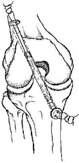
Buttons
Buttons are shown in Figure 6 and Figure 7.

Advantages
The advantages are as follows:
The Endo-button with closed loop tape is strong, if expensive.
The plastic button is cheap, available and easy to do.
Disadvantages
The disadvantages are as follows:
Fixation site is distant with increase in laxity, with the bungee cord effect.
Increased in tunnel widening.
Plastic button has low pullout strength, dependent on the sutures.
Tibial Fixation
The tibial fixation remains a problem with soft tissue graft fixation. Patients generally do not tolerate metal devices in the subcutaneous area on the front of the tibia. The interference screw gets away from that problem, but has poor performance in cyclic load. The graft tends to slip out from under the screw as the knee is cycled. A backup fixation must be used it the interference screw is used. The Intrafix (Mitek) device uses the interference screw fixation principle, but increases both the ultimate load to failure and the cyclic load performance.
Considerations
The most important consideration in ACL reconstruction is that the tunnels are put in the correct position. After this, the fixation of the graft is the next most important factor in a satisfactory clinical outcome. The physician should become proficient at one of these techniques. For revi-sions, physicians may need to have available another type of fixation to deal with hardware and tunnel expansion.
Tendon-to-Bone Healing
Studies have shown that it takes at least 8 to 12 weeks for soft tissue to heal to bone, as compared to 6 weeks for bone-to-bone healing with the patellar tendon graft. Recent studies have shown that the compression of the tendon in the tunnel with a screw speeds the time of healing, similar to internal compression in bone healing.
Donor Site Morbidity
In 1982, Lipscomb found that after harvest of the semitendinosus only the strength of the hamstrings was 102% and after harvest of both the strength was 98%. Recently, it has been shown that the internal rotation strength is decreased after the harvest of the semitendinosus. The patellofemoral pain incidence has been reported by Aligetti to be 3 to 21% after semitendinosus reconstruction. There are rare reported cases of saphenous nerve injury.
Early Rehabilitation
Prospective randomized studies by Aligetti and Marder have shown that with early and aggressive rehabilitation, there was no difference between the semitendinosus and patellar tendon grafts in stability or final knee rating. This puts to rest the argument as to whether the hamstring graft can withstand early aggressive rehabilitation protocols.
Central Quadriceps Tendon
This graft has been largely ignored in North America over the past decade. An assistant can harvest the graft while the surgeon is doing the notchplasty. It is a large diameter graft, 10 ¥ 10mm (Fig. 5.12). The tendon graft is fixed with interference screws for the bone plug and sutures tied over buttons for the tendon end. A bioabsorbable interference screw may be used at the internal aperture of the tunnel to reduce the tendon motion in the tunnel. The quadriceps tendon graft should reduce the need for the allograft or synthetic in revision cases.
 Figure. 8 The quadriceps tendon graft.
Allografts
Advantages
The allograft has no harvest site morbidity. With no harvest required, the time of the operative procedure is reduced.
Disadvantages
The main objection to the use of the allograft is the risk of disease transmission. Jackson has shown that it takes longer for the graft to incorporate and mature, meaning a longer time until the patient can return to sports. In addition, there is a limited availability of allograft materials. In the literature, Noyes has shown that in long-term follow-up, failure rates increase. In the 1997 survey of the ACL study group by Campell, none of the members used allografts for primary reconstructions.
Synthetic Grafts
The best scenario for the use of the LARS synthetic graft is when the graft can be buried in soft tissue, such as in extra-articular reconstruction. This allows for collagen ingrowth and ensures the long-term viability of the synthetic graft. It will be sure to fail early if it is laid into a joint bare, especially going around tunnel edges, and is unprotected by soft tissue.
Advantages
There is no harvest site morbidity with the use of the synthetic graft. The graft is strong from the time of initial implant. There is no risk of disease transmission.
Disadvantages
The main disadvantage is that all the long-term studies have shown high failure rate. There is the potential for reaction to the graft material with synovitis, as seen with the use of the Gore-Tex graft.With the Gore-Tex graft, there was also the increased risk of late hematogenous joint infection. The results that have been reported with the use of the Gore-Tex graft suggest that it should not be used for ACL reconstruction. Unacceptable failure rates have also been reported with the use of the Stryker Dacron ligament and the Leeds-Keio ligament. The ligament augmentation device was also found to be unnecessary.


History
The type of graft that the surgeon chooses for ACL reconstruction has evolved over the past few decades. In the 1970s, Erickson popularized the patellar tendon graft autograft that Jones had originally described in 1960. This became the most popular graft choice for the next three decades. In fact, in a survey of American Academy of Orthopaedic Surgeon members done in 2000, 80% still favored the use of the patellar tendon graft.
In the light of harvest site morbidity and postoperative stiffness associated with the patellar tendon graft, many surgeons began to look at other choices, such as semitendinosus grafts, allografts, and synthetic grafts. Fowler and then Rosenberg popularized the use of the semitendinosus. However, even Fowler was not convinced of the strength of the graft. Then, Kennedy and Fowler developed the ligament augmentation device (LAD) to supplement the semitendinosus graft. Gore-Tex (Flagstaff,AZ), Leeds-Keio, and Dacron (Stryker, Kalamazoo, MI) were choices for an alternative synthetic graft to try to avoid the morbidity of the patellar tendon graft. The initial experience was usually satisfactory, but the results gradually deteriorated with longer follow-up.
Allograft was another choice that avoided the problem of harvest site morbidity. The initial allograft that was sterilized with ethylene oxide had very poor results. Today the freeze-dried, fresh-frozen, and cryopreserved are the most popular methods of preservation of allografts.
The allograft has become a popular alternative to the autograft because it reduces the harvest site morbidity and operative time. However, Noyes has reported a 33% failure with the use of allografts for revision ACL reconstruction.
The aggressive postoperative rehabilitation program advocated by Shelbourne in the 1990s greatly diminished the problems associated with the patellar tendon graft. Before that, the patient had to be an athlete just to survive the operation and rehabilitation program. Theaggressive program emphasized no immobilization, early weight bearing, and extension exercises.
There was renewed interest in the semitendinosus during the mid-1990s. Biomechanical testing on the multiple-bundle semitendinosus and gracilis grafts demonstrated them to be stronger and stiffer than other options. This knowledge combined with improved fixation devices such as the Endo-button gave surgeons more confidence with no-bone, soft tissue grafts. The Endo-button made the procedure endoscopic, thereby eliminating the need for the second incision. Fulkerson, Staubli, and others popularized the use of the quadriceps tendon graft. This again reduced the harvest morbidity, especially when only the tendon portion was harvested.
Shelbourne has described the use of the patellar tendon autograft from the opposite knee. He claims that this divides the rehabilitation between two knees and reduces the recovery time. With the contralateral harvest technique, the average return to sports for his patients was four months. With both the patellar tendon and the semitendinosus added to the list of graft choices, the need for the use of an allograft is minimized.
The latest evolution is to use an interference fit screw to fixate the graft at the tunnel entrance. This produces a graft construct that is strong, short, and stiff. It means that the surgeon now has to learn just one technique for drilling the tunnels and can chose whatever graft he or she wishes: hamstring, patellar tendon, quadriceps tendon, or allograft.
Successful ACL reconstruction depends on a number of factors, including patient selection, surgical technique, postoperative rehabilitation, and associated secondary restraint ligamentous instability. Errors in graft selection, tunnel placement, tensioning, or fixation methods may also lead to graft failure. Comparative studies in the literature show that the outcome is almost the same regardless of the graft choice. The only significant fact from the metaanalysis, as confirmed by Yunes, is that the patellar tendon group had an 18% higher rate of return to sports at the same level. The most important aspect of the operation is to place the tunnels in the correct position. The choice of graft is really incidental. Studies by Aligetti, Marder, and O’Neill show that the only significant differences among the grafts is that the patellar tendon graft has more postoperative kneeling pain.
Evolution in Graft Choice at Carleton Sports
Medicine Clinic
The most popular graft in the early 1990s was the patellar tendon graft
(Fig. 1). With the evolution of the 4-bundle graft and improved fixation in the mid-1990s, the hamstring graft became more popular. The swing to hamstring grafts then became largely patient driven.When the patients went to therapy after the initial ACL injury, they saw how easy the rehabilitation was for the hamstring tendon and opted for that graft.
The main choices of graft for ACL reconstruction are the patellar tendon autograft, the semitendinosus autograft, and the central quadriceps tendon, allograft of patellar tendon, Achilles tendon, or tibialis anterior tendon, and the synthetic graft.

Patellar Tendon Graft
The patellar tendon graft was originally described as the gold-standard graft. It is still the most widely used ACL replacement graft (i.e., it is used in approximately 80% of cases), but it is not without problems. Shelbourne has pushed the envelope further with the patellar tendon graft. He has recently reported on the harvest of the patellar tendon graft from the opposite knee, with an average return to play of four months postoperative.
The advantages of the patellar tendon graft are early bone-to-bone healing at six weeks, consistent size and shape of the graft, and ease of Patellar Tendon Graft harvest. The disadvantages are the harvest site morbidity of patellar tendonitis, anterior knee pain, patellofemoral joint tightness with late chondromalacia, late patella fracture, late patellar tendon rupture, loss of range of motion, and injury to the infrapatellar branch of the saphenous nerve. Most of the complications are the result of the harvest of the patellar tendon.This is still the main drawback to the use of the graft.
Patellar Tendon Graft Indications
The ideal patient for an ACL reconstruction is the young, elite, competitive, pivotal athlete. This is the young athlete who wants to return to sports quickly and is going to be more aggressive in contact sports for a longer period of time. There is no upper age limit for patellar tendon reconstruction, but the younger athlete has more time to commit to knee rehabilitation. If the patellar tendon is the gold standard of grafts, then this is the graft of choice for the professional, or elite, athlete. Finally, the competitive athlete understands the value of the rehabilitation program and will not hesitate to spend three hours a day in the gym. The author’s assessment is that 50% of the success is the operation, and 50% is the rehabilitation program.
Pivoting Activities
The ACL is only required for pivotal athletics. Most nonpivotal athletes can usually cope without an ACL. Cyclists, runners, swimmers, canoeists, and kayakers, for example, can function well in their chosen sport without an intact ACL.
Athletic Lifestyle
This operation should be reserved for the athletic individual. In most activities of daily living the ACL is not essential. If the nonathlete has giving way symptoms, it is probably the result of a torn meniscus and not a torn ACL.The meniscal pathology can be treated arthroscopically, and the patient can continue with the use of a brace as necessary.
Patellar Autograft Disadvantages
Harvest Site Morbidity
The main disadvantage of the patellar tendon graft is the harvest site morbidity. The problems produced by the harvest are patellar tendonitis, quadriceps weakness, persistent tendon defect, patellar fracture, patellar tendon rupture, patellofemoral pain syndrome, patellar entrap-ment, and arthrofibrosis. The common long-term problem is kneeling pain.
Kneeling Pain
The most common complaint after patellar tendon harvest is kneeling pain. This can be reduced by harvesting through two transverse incisions. This reduces the injury to the infrapatellar branch of the saphenous nerve.
Patellar Tendonitis
Pain at the harvest site will interfere with the rehabilitation program. The strength program may have to be delayed until this settles. The problem is usually resolved in the first year, but it can prevent some high performance athletes from resuming their sport in that first year.
Quadriceps Weakness
The quads weakness may be the result of pain and the inability to participate in a strength program. If significant patellofemoral symptoms develop, the athlete may be unable to exercise the quads.
Persistent Tendon Defect
If the defect is not closed, there may be a persistent defect in the patellar tendon. This results in a weaker tendon.
Patella Entrapment
If the defect is closed too tight, the patella may be entrapped, and patellar infera may result. This will certainly result in patellofemoral pain, because of an increase in patellofemoral joint compression.
Patella Fracture
The fracture may occur during the operation or in the early postoperative period. Intraoperative patella fracture may be the result of the use of osteotomes. If the saw cuts are only 8-mm deep and 25-mm long, and the base is flat to avoid the deep V cut, an intraoperative fracture is rare. The late fractures are produced by the overruns of the saw cuts. The overruns may be prevented by cutting the proximal end in a boat shape.
The left X-ray shows a displaced transverse patellar fracture, at three months postoperative. The right X-ray shows the postoperative internal fixation with cannulated AO screws and figureof-eight wire.

Figure 2. X-ray of displaced transverse patellar fracture at three months postoperative.

Figure 3. X-ray of postoperative internal fixation with cannulated AO screws and figure-of-eight wire
Tendon Rupture
This may occur if a very large graft is taken from a small tendon. The standard is a 10-mm graft, measured with a double-bladed knife. The bone blocks are trimmed to 9 mm to make the graft passage easier.
Patellofemoral Pain
This topic is controversial in the literature. The older literature reported a high incidence of patellofemoral pain associated with ACL reconstruction. However, most of the disability could be blamed on rehabilitation programs that consisted of immobilization.There is no doubt that some patients will develop pain, some will develop crepitus, and some will have tendonitis, but results have improved with more aggressive rehabilitation programs with early motion and weight bearing. To prevent the patella from being bound down, the patella should be mobilized daily by the physiotherapist.
Arthrofibrosis
This severe problem is rarely seen now in ACL reconstructions.The true condition is idiopathic and is probably the result of fibroblastic proliferation. As a result, very little can be done to prevent it. It may be more common in the patient who forms keloid. The more common condition of loss of range of motion may be the result of incorrect tunnel placement or postoperative immobilization. In the mid-1980s, a limited range of motion hinge cast (preventing 30° of extension) was used for six weeks postoperatively, thereby causing problems in regaining extension. Many of these cases required arthroscopic debridement (10–18%, in the first year). The loss of extension was almost completely eliminated by changing to an extension splint. The acceptance of aggressive physiotherapy to regain extension eliminated the problem. This problem of postoperative stiffness made the use of a synthetic ligament, with no immobilization, very attractive. The reoperation for loss of range of motion is now very uncommon.
Tendon Rupture
This may occur if a very large graft is taken from a small tendon. The standard is a 10-mm graft, measured with a double-bladed knife. The bone blocks are trimmed to 9 mm to make the graft passage easier.
Patellofemoral Pain
This topic is controversial in the literature. The older literature reported a high incidence of patellofemoral pain associated with ACL reconstruction. However, most of the disability could be blamed on rehabilitation programs that consisted of immobilization.There is no doubt that some patients will develop pain, some will develop crepitus, and some will have tendonitis, but results have improved with more aggressive rehabilitation programs with early motion and weight bearing. To prevent the patella from being bound down, the patella should be mobilized daily by the physiotherapist.
Arthrofibrosis
This severe problem is rarely seen now in ACL reconstructions.The true condition is idiopathic and is probably the result of fibroblastic proliferation. As a result, very little can be done to prevent it. It may be more common in the patient who forms keloid. The more common condition of loss of range of motion may be the result of incorrect tunnel placement or postoperative immobilization. In the mid-1980s, a limited range of motion hinge cast (preventing 30° of extension) was used for six weeks postoperatively, thereby causing problems in regaining extension. Many of these cases required arthroscopic debridement (10–18%, in the first year). The loss of extension was almost completely eliminated by changing to an extension splint. The acceptance of aggressive physiotherapy to regain extension eliminated the problem. This problem of postoperative stiffness made the use of a synthetic ligament, with no immobilization, very attractive. The reoperation for loss of range of motion is now very uncommon..
Contraindications to Harvest of the Patellar Tendon
Preexisting Patellofemoral Pain
Is preexisting patellofemoral pain a contraindication to harvesting the patellar tendon? The conventional wisdom is yes; it would not be a wise procedure in this situation. Rather, it is like hitting a sore thumb with a hammer! In the past, when chondromalacia was seen at the time of arthroscopy, the graft choice would be changed to hamstrings.
The Small Patellar Tendon
The harvesting of the central third of the patellar tendon in a small tendon is more theoretical than practical. The advice in a small patient with a tendon width of only 25 mm would be to take a narrower graft of 8 to 9 mm or use another graft source.
Preexisting Osgoode-Schlatters Disease Shelbourne has reported that a bony ossicle from Osgoode-Schlatters disease is not a contraindication to harvest of the patellar tendon.
Because the fragment usually lies within the bony tunnel, this bone may be incorporated into the tendon graft.
Hamstring Grafts
Advantages of Hamstring Grafts
The main advantage of the hamstring graft is the low incidence of harvest site morbidity. After the harvest, the tendon has been shown by MRI to regenerate. The 4-bundle graft is usually 8mm in diameter, which is a larger cross-sectional area than the patellar tendon.
Disadvantages of Hamstring Grafts
The disadvantage of any autograft is the removal of a normal tissue to reconstruct the ACL. The harvest of the semitendinosus seems to leave the patient with minimal flexion weakness. One study did show some weakness of internal rotation of the tibia after hamstring harvest.
Injury to the saphenous nerve is rare and can be avoided with careful technique. The fixation of the graft remains one of the controversial issues.
Issues in Hamstring Grafts
The major issues with the use of hamstring grafts are:
Graft strength.
Graft fixation.
Graft healing.
Donor site morbidity.
Early rehabilitation.
Graft strength and stiffness.
In one of the earlier studies, Noyes reported that one strand of the semi-t was only 70% the strength of the ACL. However, hecompared this to a 15-mm-wide patellar tendon graft that was 125% the strength of the native ACL. This was widely quoted as a reason to use the patellar tendon graft rather than the hamstring.With the advent of the multiple bundles of hamstrings, this graft now has twice the strength of the native ACL (Fig. 7). Sepaga later reported that the semitendinosus and gracilis composite graft is equal to an 11-mm patellar tendon graft. Marder and Larson felt that if all the bundles are equally tensioned, the double-looped semi-t and gracilis is 250% the strength of the normal ACL. Hamner, however, emphasized that the strength is only additive if the bundles are equally tensioned.
Soft Tissue Fixation Techniques
There are various techniques for securing the soft tissue to the bony tunnel in ACL reconstruction. Each one has strengths and weaknesses. Pinczewski pioneered the use of the RCI interference fit metal screw for soft tissue fixation. The use of a similar type of bioabsorbable screw that was used in bone tendon bone fixation was a natural evolution. To overcome the weak fixation in poor quality bone, the use of a round pearl, made of PLLA or bone, was developed.This improved the pullout strength by 50%.The Endo-button, popularized by Tom Rosenberg, was improved with the use of a continuous polyester tape. This made the fixation stronger and avoided the problems of tying a secure knot in the tape. The cross-pin fixation has proven to be the strongest, but has a significant fiddle factor to loop the tendons around the post. The Arthrex technique is the easiest to use.Weiler, Caborn, and colleagues have summarized the current concepts of soft tissue fixation. The estimates of the force on the normal ACL during activities of daily living are as follows:
Level walking: 169N
Ascending stairs: 67N
Descending stairs: 445N
Ascending ramp: 27N
Descending ramp: 93N
It is commonly quoted that a person needs more than 445N pullout strength of the device just to handle the activities of daily living. However, Shelbourne has reported good results with the patellar tendon graft fixed by tying the leader sutures over periosteal buttons (Ethicon, J&J, Boston, MA). This form of fixation has a low failure strength, but is clinically successful. The gold standard of the interference fit screw fixation of the bone tendon bone, 350 to 750N, has been used to compare the soft tissue fixation.
The pullout strengths also vary from tibia to the femur. The femoral pullout is higher because the tunnel is angled to the graft and the pull is against the screw that is placed endoscopically. In the tibial tunnel, the graft pulls away from the screw in the direct line of the tunnel.
The initial fixation points were at a distance from the normal anatomical fixation of the ACL. The trend has been to move the fixation closer to the internal aperture of the tunnel. This shortening of the intraarticular length has improved the stiffness of the graft.
The pullout strength of bioabsorbable screw can vary widely depending on its composition. The screw fixation has also been shown to be bone quality dependent. These considerations should be taken into account when choosing a femoral fixation device for soft tissue grafts.
Disadvantages
The disadvantages of the hamstring graft are the various methods used to fix the graft to bone, including staples, Endo-button, and interference fit screws. Furthermore, the graft harvest can be difficult, the tendons can be cut off short, and there is a longer time for graft healing to bone, approximately 10 to 12 weeks.
Pullout Strengths of Soft Tissue Devices
The fixation of the graft depends on both the tibial and femoral fixation. The rehabilitation protocol should reflect the type of fixation used. All the femoral fixation devices provide reasonable fixation. The cyclic load is more important than the ultimate load to failure. The interference screw fares worst with cyclic loads.
Interference Fit Screws
The interference fit screw is shown is Figure 4.

Figure 4. The interference screw fixation of the soft tissue graft in a cadaver model
.
Advantages
The advantages are as follows:
Quick, familiar, and easy to use.
Direct bone to tendon healing, with Sharpey’s fibers at the tunnel aperture.
Less tunnel enlargement.
Disadvantages
The disadvantages are as follows:
Longer graft preparation time.
Bone quality dependent.
Damage to the graft with the screw.
Divergent screw has poor fixation.
Removal of metal screw makes revision difficult.
Several refinements have been made to the interference screw technique to increase the pullout strength and cyclic load performance. The end of the graft may be backed up with a round ball of PLLA, the Endo- Pearl (Linvatec, Largo, FL) or bone to abut against the screw and prevent the slippage of the graft under the screw. The tunnels may be dilated or compacted when the bone is osteopenic.A longer screw with a heavy whipstitch in the graft improves pullout strength. The leader sutures from the graft may be tied over a button or post on the tibial side to back up the screw fixation.
Cross-Pin Fixation
The cross-pin fixation is shown in Figure 5

Figure.5. The Arthrex transfix pin fixation of soft tissues.
Advantages
The advantages are as follows:
Strongest tested fixation.
May individually tension all bundles of graft.
Disadvantages
The disadvantages are as follows:
Pin may tilt in soft bone and lose fixation.
Steep learning curve of fiddle factor.
Special guides are required.
Buttons
Buttons are shown in Figure 6 and Figure 7.

Figure 6. The Endo-button periosteal cortical femoral fixation of hamstring grafts.

Figure 7 The periosteal button fixation of soft tissue grafts.
Advantages
The advantages are as follows:
The Endo-button with closed loop tape is strong, if expensive.
The plastic button is cheap, available and easy to do.
Disadvantages
The disadvantages are as follows:
Fixation site is distant with increase in laxity, with the bungee cord effect.
Increased in tunnel widening.
Plastic button has low pullout strength, dependent on the sutures.
Tibial Fixation
The tibial fixation remains a problem with soft tissue graft fixation. Patients generally do not tolerate metal devices in the subcutaneous area on the front of the tibia. The interference screw gets away from that problem, but has poor performance in cyclic load. The graft tends to slip out from under the screw as the knee is cycled. A backup fixation must be used it the interference screw is used. The Intrafix (Mitek) device uses the interference screw fixation principle, but increases both the ultimate load to failure and the cyclic load performance.
Considerations
The most important consideration in ACL reconstruction is that the tunnels are put in the correct position. After this, the fixation of the graft is the next most important factor in a satisfactory clinical outcome. The physician should become proficient at one of these techniques. For revi-sions, physicians may need to have available another type of fixation to deal with hardware and tunnel expansion.
Tendon-to-Bone Healing
Studies have shown that it takes at least 8 to 12 weeks for soft tissue to heal to bone, as compared to 6 weeks for bone-to-bone healing with the patellar tendon graft. Recent studies have shown that the compression of the tendon in the tunnel with a screw speeds the time of healing, similar to internal compression in bone healing.
Donor Site Morbidity
In 1982, Lipscomb found that after harvest of the semitendinosus only the strength of the hamstrings was 102% and after harvest of both the strength was 98%. Recently, it has been shown that the internal rotation strength is decreased after the harvest of the semitendinosus. The patellofemoral pain incidence has been reported by Aligetti to be 3 to 21% after semitendinosus reconstruction. There are rare reported cases of saphenous nerve injury.
Early Rehabilitation
Prospective randomized studies by Aligetti and Marder have shown that with early and aggressive rehabilitation, there was no difference between the semitendinosus and patellar tendon grafts in stability or final knee rating. This puts to rest the argument as to whether the hamstring graft can withstand early aggressive rehabilitation protocols.
Central Quadriceps Tendon
This graft has been largely ignored in North America over the past decade. An assistant can harvest the graft while the surgeon is doing the notchplasty. It is a large diameter graft, 10 ¥ 10mm (Fig. 5.12). The tendon graft is fixed with interference screws for the bone plug and sutures tied over buttons for the tendon end. A bioabsorbable interference screw may be used at the internal aperture of the tunnel to reduce the tendon motion in the tunnel. The quadriceps tendon graft should reduce the need for the allograft or synthetic in revision cases.

Allografts
Advantages
The allograft has no harvest site morbidity. With no harvest required, the time of the operative procedure is reduced.
Disadvantages
The main objection to the use of the allograft is the risk of disease transmission. Jackson has shown that it takes longer for the graft to incorporate and mature, meaning a longer time until the patient can return to sports. In addition, there is a limited availability of allograft materials. In the literature, Noyes has shown that in long-term follow-up, failure rates increase. In the 1997 survey of the ACL study group by Campell, none of the members used allografts for primary reconstructions.
Synthetic Grafts
The best scenario for the use of the LARS synthetic graft is when the graft can be buried in soft tissue, such as in extra-articular reconstruction. This allows for collagen ingrowth and ensures the long-term viability of the synthetic graft. It will be sure to fail early if it is laid into a joint bare, especially going around tunnel edges, and is unprotected by soft tissue.
Advantages
There is no harvest site morbidity with the use of the synthetic graft. The graft is strong from the time of initial implant. There is no risk of disease transmission.
Disadvantages
The main disadvantage is that all the long-term studies have shown high failure rate. There is the potential for reaction to the graft material with synovitis, as seen with the use of the Gore-Tex graft.With the Gore-Tex graft, there was also the increased risk of late hematogenous joint infection. The results that have been reported with the use of the Gore-Tex graft suggest that it should not be used for ACL reconstruction. Unacceptable failure rates have also been reported with the use of the Stryker Dacron ligament and the Leeds-Keio ligament. The ligament augmentation device was also found to be unnecessary.

Figure 9 The insertion of the BioScrew through the anteromedial portal

Figure 10. The insertion of the BioScrew into the femoral tunnel
Subscrever:
Mensagens (Atom)