Pesquisar neste blogue

sábado, 9 de julho de 2016

Physical Inactivity Is Deadly! Strategies For Physiotherapists To Get Patients Active

http://www.physio-pedia.com/conferences/ifompt/

This Parallel Session from Day 4 of IFOMPT 2016 was entitled: Physical Inactivity Is Deadly! Strategies For Physiotherapists To Get Patients Active. The provided Learning Objectives were as follows:
  • Understand epidemiologic and public health issues associated with physical inactivity, and discuss the public health role of musculoskeletal physiotherapists in primary prevention of non-communicable diseases
  • Measure physical activity and identify barriers to patient adherence to physical activity participation
  • Implement interventions to enable/support patients in increasing their physical activity and decreasing sedentary behaviour
The full abstract can be found here.
This session had what I thought was a surprisingly low turnout given that this topic is so pertinent to many of our patients; as Dr Debra Shirley noted during her introduction, many MSK patients are at risk of comorbidities such as noncommunicable disease (NCD) therefore addressing the risk of these NCDs through physical activity education should be a cornerstone of physiotherapy. As she also commented, however, there were other interesting sessions in the same time slot as always happens at content-packed conferences such as IFOMPT, therefore it is difficult for delegates to choose between them.
IMG_20160707_060859 cropped and rotated
Dr Shirley first posed the question: “are adults sufficiently active?” The above map shows countries that have the lowest proportions of inhabitants meeting physical activity guidelines in darker colours (apologies that the photo is a little blurry). Dr Shirley pointed out that some improvement has occurred compared to a few years ago when 2/3 of the world’s population were insufficiently active but even with this positive change, there is room for further improvement. She also provided some data from the World Health Organization which has found that in general terms, women are more inactive than men and that the Americas and Eastern Europe are the least active areas.
Dr Shirley then went on to note that physical activity has been shown to reduce the risk of mortality due to NCDs. Inactivity is the 4th highest risk factor for death after hypertension, tobacco use and high blood glucose. There are many known health benefits of physical activity which can be seen in the slide below. Despite the obvious advantages of being physically active, we have just seen in the above map that people are still not active enough. As Dr Shirley commented, if only it was as easy as getting patients to take a pill!


The Many Health Benefits of Physical Activity
Risk of Disease Relative to Amount of Physical Activity
Health Benefits of Physical Activity
The Many Health Benefits of Physical Activity

She then asked: “Who should be responsible for getting patients more active?” She shared a slide listing the seven best investments in physical activity. Number four on this list was integration of physical activity and NCD prevention into primary health care systems. She also showed a position statement from the World Confederation for Physical Therapy that included promoting health and emphasising the importance of physical activity within the scope of physiotherapy. Dr Shirley noted that health care professionals can reach and influence large numbers of people in the population for the following reasons;
  • 70-80% of adults visit their GP each year in developed countries.
  • Patients are often inactive and at risk of NCDs
  • Patients consider primary care professionals appropriate to discuss health promotion with
  • Physical activity interventions based in primary care can potentially generate larger health gains
Dr Shirley shared the estimated number of occasions of service in physiotherapy private practice in Australia in 2008 at 15.6 million* according to statistics from the Australian Physiotherapy Association. Clearly these numbers show that there is a huge number of opportunities to discuss physical activity with patients.
(*Please note that this number is awaiting confirmation from Dr Shirley)
Dr Shirley encouraged audience participation by asking us “What are the Physical Activity guidelines?” and had us respond through www.socrative.com. She was encouraged to see that a lot of people answered along the right lines.
When Professor Dean started speaking, she mentioned again the draw of the other parallel sessions running at the same time as the Physical Inactivity one. She said that as manual physiotherapists we obviously embrace learning about manual techniques but she also noted that things have changed and that we clearly need to redirect our practice to expand our scope of practice to include physical behaviour change.


IMG_20160707_062536 cropped and rotated
Professor Dean and the Activity Pyramid

Professor Dean noted that even if you follow the physical activity guidelines but are sedentary the remainder of the time, you’re still at risk of NCD. She also rhetorically asked, “how much sitting is ok?”  and went on to report that humans were not designed to sit, not from endocrine, cardiopulmonary or biomechanical perspectives. She discussed changes that are being implemented in some workplaces to combat excessive sitting such as walking a minimum of five minutes every hour or the use of standing desks. Wearable technology such as pedometers are also used to encourage more activity. 10,000 steps per day are considered in the active range while < 5000 steps per day is a criterion for sedentary lifestyle; most of us average 6000-65oo steps per day.
Professor Dean discussed the notion of micro-exercise and how small bouts of moderate physical activity add up. She gave the example of Stockholm Airport where you can charge your own electronic devices using cycle ergometers. More examples of micro-exercises can be seen in the slide below.
IMG_20160707_062829 cropped and rotated
Professional Dean also discussed some tools that can be used for tracking and promoting physical activity. These can be seen in the slide below, although she noted that observations are obviously not practical in a clinical setting. Assessment tools and other resources that were discussed included;
Professor Ann Moore first joked that one of the health behaviour questions Professor Dean should have asked was: “Have you attended an IFOMPT conference, sat through all the sessions, booked a hotel right next to the conference and eaten a highly calorific meal every day for five days?!” Indeed. I admittedly felt pretty slovenly at that moment in time.
Professor Moore emphasized the idea of making every consultation count by tailoring interventions to each patient, creating individualized self-management strategies to support the interventions, providing advice on weight loss etc and providing sufficient education, advice and discussion as needed to ensure the full understanding of both parties. The latter point is particularly important when it comes to discussions of self-management. Professor Moore noted that at least in England, there is pressure to see patients for fewer visits and a shorter amount of time under the NHS. She said there was a maximum of 1.5 treatments available for patients with low back pain and she pointed out that so few visits would clearly be insufficient for setting up and supporting a good self-management program. Printing exercises off a website and giving the patient a handout simply doesn’t work and she said she found it worrying if this was considered reasonable. In contrast, a well-structured and patient-centred self-management program will benefit patients and their families, health care professionals, the NHS (or other health care system) as well as the government and policy makers .


Benefits of Self-Management Program

One major challenge of self-management is that there is often a difference in understanding of self-management between health care professionals and patients. A recent study found different viewpoints on this matter is summarized below.  .


Professor Moore and Different Viewpoints of Self-Management

Clearly, if some of these negative viewpoints are possible, and particularly if a therapist does not educate the patient about what self-management is relative to what the patient thinks self-management is, then the management program won’t work. Patients must understand that you’re not “abandoning” them and this can be done by leaving more time available for discussion in the first session and also by making more information available for patients about their condition and treatment.
In the final portion of the session, Dr Shirley discussed implementation of interventions. Various formats can be utilized for physical activity education and are overviewed in the following slide. All have been shown to be effective but none have been shown to be superior over other formats.
IMG_20160707_070033 cropped and rotated
Dr Shirley also discussed the sequence of promotion in primary care by Stevenson and Roach (2012) which is shown below.


Sequence of Promotion in Primary Care

Again, knowing that any behaviour change can be difficult to effect, Dr Shirley discussed the NICE guidance on Behaviour Change and the stages of change (Precontemplation, Contemplation, Preparation, Action and Maintenance). She noted that we need to focus on patients in the first three categories using education, finding/removing barriers and motivation. Many perceived barriers prevent people from forming new positive behaviours such as physical activity, including work commitments, lack of leisure time, caring for children or older people, lack of money, poor health and lack of motivation. There are many suggestions for removing these barriers (see below) and Dr Shirley noted that a key point to emphasize is that being physically active does not have to cost much money.
IMG_20160707_070605 cropped and rotated
Additional strategies for increasing physical activity levels at home, at work and while traveling can be found below. Dr Shirley noted that over 1/3 of Australian households own a dog and likened a dog to a living breathing physical activity reminder. So many of these strategies sound like common sense and yet it is amazing how few people actually apply them.


IMG_20160707_070754 cropped and rotated
Strategies for Increasing Physical Activity at Home
IMG_20160707_070808 cropped and rotated
Strategies for Increasing Physical Activity at Work
IMG_20160707_070936 cropped and rotated
Strategies for Increasing Physical Activity while Traveling

To finish her part of the session, Dr Shirley mentioned various resources available for the promotion and adoption of a physically active lifestyle. These included;
IMG_20160707_071345 cropped and rotated
The Implication/Conclusions for this session were listed as: Physiotherapists are ideally positioned to incorporate physical activity advice for health promotion into practice. Evidence-based approaches and tools that are amenable to integration into contemporary physiotherapy practices need to be exploited in every patient as indicated, and incorporated into entry-to-practice physiotherapy professional education. In this way, physiotherapists could have a major impact on preventing/reversing inactivity as a major public health issue.
IMG_20160707_065758 rotated and cropped


Personally, I have attended conference sessions on Physical Activity before and I admittedly am still not bringing up the subject with the majority of my patients. There is a research finding from many years ago indicating that on average, people need to be told about the benefits of a certain behaviour change three times before they actually get serious about making the change. Perhaps it is similar for physiotherapists adapting changes in their clinical practice. For me, I’ve listened to speakers on this topic enough times that I’m at the stage where I need to get serious about making this change and this session was a great springboard for that.

The Nine Mental Skills of Successful Athletes

www.sportpsych.org/nine-mental-skills-overview

Autor: Jack J. Lesyk, Ph.D.

You don’t have to be a professional athlete or an Olympic champion to be a successful athlete. Nor do you have to have a room full of trophies, win a state championship, or make the front page of the sports section. Successful athletes that I’ve worked with include an eleven year-old figure skater who has not yet won a competition, a high school golfer with a zero handicap, a middle-aged runner whose goal is to complete her first marathon, a weight lifter who holds several world records, and an Olympic medalist.
What these athletes have in common is that their sport is important to them and they’re committed to being the best that they can be within the scope of their limitations – other life commitments, finances, time, and their natural ability. They set high, realistic goals for themselves and train and play hard. They are successful because they are pursuing their goals and enjoying their sport. Their sport participation enriches their lives and they believe that what they get back is worth what they put into their sport.
There are nine, specific mental skills that contribute to success in sports. They are all learned and can be improved with instruction and practice. At the Ohio Center for Sport Psychology we work with serious athletes of all ages and ability levels to help them learn and sharpen these important skills.
We believe that our work is worthwhile because the same mental skills that athletes use in achieving success in sports can be used to achieve success in other areas of their lives.

A Brief List of the Nine Mental Skills

Successful Athletes:

  1. Choose and maintain a positive attitude.
  2. Maintain a high level of self-motivation.
  3. Set high, realistic goals.
  4. Deal effectively with people.
  5. Use positive self-talk.
  6. Use positive mental imagery.
  7. Manage anxiety effectively.
  8. Manage their emotions effectively.
  9. Maintain concentration.

Mental Skills Training

These nine mental skills are necessary for performing well in sport as well as in non-sport performance situations. At the Ohio Center for Sport Psychology:
  • We believe that these skills are learned and can be improved through instruction and practice.
  • We begin our work with each individual by assessing his current proficiency in each of the skills.
  • We develop a plan for teaching and enhancing the specific skills that need improvement for the individual.
  • We periodically reassess the client’s proficiency in each of the skills in order to evaluate our progress.

The Performance Pyramid

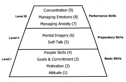
Although each of the nine skills is important, its primary importance will occur during one of three phases: long-term development, immediate preparation for performance, and during performance itself.
Level I - These mental skills constitute a broad base for attaining long-term goals, learning, and sustaining daily practice. They are needed on a day-by-day basis for long periods of time, often months and years.
Level II - These skills are used immediately before performance to prepare for performance. They maybe used just before competition begins, or immediately before a specific performance action, such as a golf shot or a free throw in basketball.
Level III - These skills are used during actual performance behavior.
The pyramid below represents the relationship of the nine skills to one another. Each of the higher levels incorporates and is based upon the skills of the preceding levels.

Detailed Descriptions of the Nine Mental Skills

1. Attitude

Successful athletes:
  • Realize that attitude is a choice.
  • Choose an attitude that is predominately positive.
  • View their sport as an opportunity to compete against themselves and learn from their successes and failures.
  • Pursue excellence, not perfection, and realize that they, as well as their coaches, teammates, officials, and others are not perfect.
  • Maintain balance and perspective between their sport and the rest of their lives.
  • Respect their sport, other participants, coaches, officials, and themselves.

2. Motivation

Successful athletes:
  • Are aware of the rewards and benefits that they expect to experience through their sports participation.
  • Are able to persist through difficult tasks and difficult times, even when these rewards and benefits are not immediately forthcoming.
  • Realize that many of the benefits come from their participation, not the outcome.

3. Goals and Commitment

Successful athletes:
  • Set long-term and short-term goals that are realistic, measurable, and time-oriented.
  • Are aware of their current performance levels and are able to develop specific, detailed plans for attaining their goals.
  • Are highly committed to their goals and to carrying out the daily demands of their training programs.

4. People Skills

Successful athletes:
  • Realize that they are part of a larger system that includes their families, friends, teammates, coaches, and others.
  • When appropriate, communicate their thoughts, feelings, and needs to these people and listen to them as well.
  • Have learned effective skills for dealing with conflict, difficult opponents, and other people when they are negative or oppositional.

5. Self-Talk

Successful athletes:
  • Maintain their self-confidence during difficult times with realistic, positive self-talk.
  • Talk to themselves the way they would talk to their own best friend
  • Use self-talk to regulate thoughts, feelings and behaviors during competition.

6. Mental Imagery

Successful athletes:
  • Prepare themselves for competition by imagining themselves performing well in competition.
  • Create and use mental images that are detailed, specific, and realistic.
  • Use imagery during competition to prepare for action and recover from errors and poor performances.

7. Dealing Effectively with Anxiety

Successful athletes:
  • Accept anxiety as part of sport.
  • Realize that some degree of anxiety can help them perform well.
  • Know how to reduce anxiety when it becomes too strong, without losing their intensity.

8. Dealing Effectively with Emotions

Successful athletes:
  • Accept strong emotions such as excitement, anger, and disappointment as part of the sport experience.
  • Are able to use these emotions to improve, rather than interfere with high level performance

9. Concentration

Successful athletes:
  • Know what they must pay attention to during each game or sport situation.
  • Have learned how to maintain focus and resist distractions, whether they come from the environment or from within themselves.
  • Are able to regain their focus when concentration is lost during competition.
  • Have learned how to play in the “here-and-now”, without regard to either past or anticipated future events.

Application of the Nine Mental Skills to Non-sport Performance Situations

The nine mental skills associated with athletic success are the same mental skills associated with performance in a wide variety of non-sport, performance situations. Let’s take a look at some of these.

Characteristics of A Performance Situation:

  • The situation is often scheduled or anticipated in advance.
  • The situation usually has a defined beginning and an end.
  • The circumstances are known in advance.
  • The rules and constraints are known in advance.
  • The results are evaluated by standards (or natural consequences) that are usually known in advance.
  • The results are uncertain and may involve psychological risk and/or danger.
  • The results are important to the performer.
  • The performer’s behavior is goal-oriented.
  • The results are influenced by the performer’s skillful behavior

Examples of Performance Situations

  • An important job interview
  • Performing a solo with a symphony orchestra
  • Auditioning for a role in a drama production
  • Giving a class presentation
  • Taking a driver’s examination
  • Giving a talk to the PTA
  • Testifying in court
  • Taking the state medical exam
  • Performing brain surgery
  • Landing an airplane
  • A firefighter entering a burning building
  • Participating in a military or police attack
  • An astronaut landing a vehicle on the surface of the moon
  • Rock Climbing

domingo, 1 de maio de 2016

FIFA Diploma in Football Medicine - FREE online course!


The FIFA Diploma in Football Medicine is a FREE online course, designed to help clinicians learn how to diagnose and manage common football-related injuries and illnesses. The course is designed specifically for primary care clinicians including doctors, physiotherapist and other allied health practitioners. 
http://f-marc.com/footballdiploma/


quarta-feira, 27 de abril de 2016

MRI OF THE ATHLETE WITH ELBOW PAIN

 http://www.aspetar.com/journal/viewarticle.aspx?id=16#.VyEvczArK01

– Written by Richard Kijowski, USA

INTRODUCTION
MRI is commonly used to evaluate athletes with elbow pain. MRI can acquire high resolution images with excellent tissue contrast in multiple planes.  It is therefore well-suited for assessing the bones, ligaments, tendons and nerves of the elbow which may all be sources of pain in athletes. MRI is particularly helpful for evaluating athletes with non-localisable elbow pain and athletes with a history of acute trauma to the elbow superimposed on chronic injury from repetitive micro-trauma. Even when the cause of elbow pain in athletes can be diagnosed clinically, MRI can assess the severity of the injury and thereby provides useful information for determining the most appropriate treatment option and estimating recovery time. This article will review the role of MRI in evaluating a wide spectrum of elbow injuries in athletes.

OSSEOUS INJURIES
Osteochondral lesions
Osteochondral lesions comprise the vast majority of osseous injuries in young athletes. Osteochondral lesions typically occur in adolescent baseball pitchers, tennis and squash players and gymnasts who have a history of chronic repetitive micro-trauma to the elbow. Most osteochondral lesions are located within the anterolateral capitellum although there are rare reports of lesions involving the trochlea, radial head and olecranon. Although the exact aetiology of osteochondral lesions is currently unknown, most authors believe that the condition is primarily due to chronic repetitive micro-trauma to the poorly vascularised capitellum. Athletes with osteochondral lesions typically present with pain, tenderness and swelling over the lateral aspect of the elbow. Radiographs may show rarefaction, non-displaced fragmentation or focal defect of the subchondral bone of the capitellum1.
MRI is commonly used to evaluate athletes with osteochondral lesions of the elbow. MRI is superior to radiographs for detecting early osseous injuries and can determine the exact size and location of the osteochondral lesion. MRI can also be used to assess the stability of the osteochondral lesion which is the single most important prognostic factor that determines its likelihood to heal with conservative therapy. Osteochondral lesions have variable signal intensity on T1-weighted and T2-weighted images. Stable lesions are surrounded by patchy bone marrow oedema or show no surrounding T2 signal abnormality. Unstable lesions are surrounded by a high T2-signal rim or cysts (Figure 1). Displacement of an unstable osteochondral lesion may lead to a focal defect within the articular surface with an associated loose body within the elbow joint1.

NON-DISPLACED FRACTURES
MRI is commonly used to detect and characterise non-displaced fractures in athletes. MRI is especially useful for detecting non-displaced radial head fractures following acute injury and avulsion fractures of the sublime tubercle due to chronic repetitive micro-trauma which are often difficult to identify on radiographs. MRI can also detect ‘little leaguers elbow’, a repetitive stress injury of the physis at the medial epicondyle apophysis, during the early stage of the injury before radiographic changes become apparent. MRI is also useful for identifying and assessing the severity of stress injuries to the olecranon process and other bones of the elbow which are especially common in elite baseball pitchers who generate high forces across the elbow joint during the throwing motion (Figure 2)2.

LIGAMENT INJURIES
Ulnar collateral ligament tears
Tears of the ulnar collateral ligament in athletes may occur following acute valgus injury but are more commonly the result of chronic repetitive stress to the elbow elicited by overhead throwing activities. Most athletes with ulnar collateral ligament tears present with medial elbow pain which is most severe during the acceleration phase of the throwing motion. Individuals may also describe a sensation of elbow ‘opening’ while throwing or decreased ability to maximise velocity or distance during the throwing motion. Most tears of the ulnar collateral ligament involve the anterior bundle which is the primary stabiliser of the elbow joint against valgus and internal rotatory stress. Most anterior bundle tears involve the mid-portion of the ligament although complete soft tissue avulsion of the proximal and distal attachments may also occur. Partial thickness tears of the deep fibre of the distal attachment of the anterior bundle have also been described and are especially common in elite baseball pitchers3.
MRI may be used to evaluate the ulnar collateral ligament in athletes. MRI has 100% sensitivity for detecting full-thickness tears but only 57% sensitivity for detecting partial-thickness tears of the anterior bundle of the ulnar collateral ligament. The normal anterior bundle of the ulnar collateral ligament appears as a thin band of low signal intensity on coronal T1-weighted and T2-weighted images that extends along the medial joint line of the elbow. The MRI findings of a torn anterior bundle of the ulnar collateral ligament include redundancy, irregularity and poor definition of the ligament and abnormal signal intensity within and surrounding the ligament (Figure 3). The abnormal morphology and signal intensity of the torn ligament is thought to be due to the presence of oedema and haemorrhage. Chronic tears of the anterior bundle of the ulnar collateral ligament are often difficult to detect on MRI due the absence of surrounding soft tissue oedema and haemorrhage3.

MR arthrography of the elbow using saline or gadolinium intra-articular contrast can also be used to evaluate the ulnar collateral ligament in athletes. The presence of intra-articular contrast allows for better delineation of the under surface of the anterior bundle of the ulnar collateral ligament and significantly improves the sensitivity for detecting partial-thickness tears. On MR arthrography, the distal insertion of the anterior bundle attaches within 1 mm of the articular margin of the coronoid process of the ulna. Any intra-articular contrast extending below the attachment of the anterior bundle to the base of the coronoid process is abnormal and indicates a partial-thickness tear of the ulnar collateral ligament (Figure 4). A full-thickness tear of the ulnar collateral ligament results in contrast extravasation into the medial soft tissue of the elbow. This finding may be especially useful for detecting chronic tears of the ulnar collateral ligament4.

Valgus extension overload syndrome
Valgus extension overload syndrome is a condition seen in overhead throwing athletes who have an intact but attenuated ulnar collateral ligament due to chronic repetitive micro-trauma. Attenuation of the anterior bundle of the ulnar collateral ligament allows the posteromedial olecranon tip to impinge against the medial aspect of the olecranon fossa during the throwing motion which results in formation of olecranon tip osteophytes and damage to the articular cartilage of the posteromedial trochlea. Subtle valgus laxity may also lead to excessive tension on the medial soft tissue of the elbow which may cause chronic repetitive injury to the common flexor tendon origin and ulnar nerve. Athletes with valgus extension overload syndrome typically present with chronic vague posteromedial elbow pain. MRI may be useful for diagnosis and for determining the extent of injury to the ulnar collateral ligament, trochlear cartilage, common flexor tendon origin and ulnar nerve5.

Lateral collateral ligament tears
Tears of the lateral collateral ligament are relatively uncommon in athletes and are typically caused by an acute varus extension injury to the elbow. Lateral collateral ligament injuries primarily consist of full-thickness tears of the proximal attachment of the ulnar band of the lateral collateral ligament which is the primary stabiliser of the elbow joint against varus and external rotatory stress. Tears of the ulnar band of the lateral collateral ligament result in posterolateral rotatory instability of the elbow. Athletes with posterolateral rotatory instability usually present with a long history of lateral elbow pain and a catching or snapping sensation during movement of the elbow. MRI plays an important role in diagnosis since the condition is often difficult to detect using physical examination and radiographs. The normal ulnar band of the lateral collateral ligament appears as a thin band of low signal intensity on coronal T1-weighted and T2-weighted images that extends along the lateral joint line of the elbow and then crosses posterior to the radial head to insert onto the proximal ulna. The MRI findings of a torn lateral collateral ligament include redundancy, irregularity and poor definition of the ligament and abnormal signal intensity within and surrounding the ligament (Figure 5)6.

TENDON INJURIES
Lateral epicondylitis
Lateral epicondylitis is a pathologic condition of the common extensor tendon at its origin from the lateral epicondyle. The condition is primarily seen in athletes between 40 and 60 years of age. Lateral epicondylitis is also known as tennis elbow since over 50% of tennis players develop the condition at one time or another. Athletes with lateral epicondylitis present with chronic lateral elbow pain exacerbated by activities that require resisted extension of the wrist. The condition is thought to represent an incomplete healing response to an initial microscopic or macroscopic avulsion injury of the common extensor tendon origin. Lateral epicondylitis typically involves the tendon origins of the extensor carpi radialis brevis muscle and occasionally extensor digitorum communis and extensor carpi radialis longus muscles from the lateral epicondyle7.
Most athletes with lateral epicondylitis are easily diagnosed using clinical history and physical examination. However, MRI may be useful for determining the extent of tissue damage to the common extensor tendon origin and to exclude other causes of lateral elbow pain in patients who do not respond well to conservative treatment. The normal common extensor tendon origin shows homogenous low signal intensity on both T1-weighted and T2-weighted images. The common extensor tendon origin in athletes with lateral epicondylitis is usually thickened and shows increased T1 and T2 signal intensity. In individuals with severe lateral epicondylis, the common extensor tendon origin may be thinned and show areas of intense fluid-like signal intensity on T2-weighted images (Figure 6). Additional MRI findings include bone marrow oedema of the lateral epicondyle, anconeus muscle oedema and fluid within the radial head bursa7.

Medial epicondylitis
Medial epicondylitis is a pathologic condition of the common flexor tendon at its origin from the medial epicondyle and is much less common than lateral epicondylitis. The condition is typically seen in athletes between 40 and 60 years of age who are involved in sports activities which generate repetitive valgus and flexion forces at the elbow. Athletes with lateral epicondylitis present with chronic medial elbow pain exacerbated by activities that require resisted flexion of the wrist and pronation of the forearm. The condition is thought to represent an incomplete healing response to an initial microscopic or macroscopic avulsion injury of the common flexor tendon. Medial epicondylitis most commonly involves the tendon origins of the flexor carpi radialis and pronator teres muscles from the medial epicondyle. On MRI, the origin of the common flexor tendon in athletes with medial epicondylitis is usually thickened and shows increased signal intensity on both T1-weighted and T2-weighted images. However, the common flexor tendon origin may be thinned and show areas of intense fluid-like signal intensity on T2-weighted images especially in the advanced stages of the disease process (Figure 7). In contrast, the normal common flexor tendon origin shows homogenous low signal on both T1-weighted and T2-weighted images8.

Biceps tendon injuries
Distal biceps tendon tears most commonly occur in the dominant extremity of male athletes between 40 and 60 years of age. Tears of the biceps tendon may be partial or complete and typically occur at the insertion site of the tendon into the radial tuberosity. Biceps tendon tears are almost always caused by a single traumatic event in which a sudden extension force is applied to the arm with the elbow flexed 90°. Athletes usually present with an acute onset of pain and swelling in the antecubital fossa of the elbow following a single traumatic event. The exact aetiology of distal biceps tendon tear is currently unknown. However, degenerative factors are thought to play a major role in the pathogenesis as almost all torn biceps tendons show evidence of tendinopathy on histopathologic analysis9.
MRI may be useful for evaluating athletes with distal biceps tendon tears, especially individuals with partially torn or completely torn but non-retracted tendons which are often difficult to clinically diagnose. The distal biceps tendon is best evaluated on axial T1-weighted and T2-weighted images. Complete tendon tear is characterised by the absence of the low signal intensity distal biceps tendon at its insertion site on the radial tuberosity and by the presence of soft tissue oedema within the antecubital fossa. A variable amount of retraction of the distal biceps tendon is usually noted on sagittal images (Figure 8). Partial tendon tear is characterised by the presence of increased signal intensity within an abnormally thickened or thinned distal biceps tendon. Secondary findings of a partially torn distal biceps tendon include bone marrow oedema within the radial tuberosity and fluid within the bicipitoradial bursa9.

Triceps tendon injury
Triceps tendon tears are a rare injury in athletes and occur in both males and females and in individuals of all ages. Tear of the triceps tendon is almost always the result of a single traumatic event such as a fall on an outstretched hand, a direct blow to the posterior elbow or a forceful eccentric contraction of the triceps muscle with the elbow flexed. Most tears occur at the insertion site of the triceps tendon into the olecranon process of the proximal ulna. Individuals with triceps tendon tear usually present with an acute onset of pain and swelling in the posterior elbow following a single traumatic even. MRI may be useful for distinguishing between partial and complete tear of the triceps tendon and for determining the amount of retraction of a completely torn tendon. The triceps tendon is best visualised on sagittal T1-weighted and T2-weighted images. Partial tendon tear is characterised by irregular contour and increased signal intensity within the distal triceps tendon with oedema within the surrounding subcutaneous soft tissue (Figure 9). Complete tear of the triceps tendon is characterised by a large fluid filled gap between the distal triceps tendon and the olecranon process with a large amount of surrounding soft tissue oedema10.

NERVE INJURIES
Athletes place forceful repetitive stress on the elbow joint which results in chronic traction and compression of the ulnar, radial and median nerves. Compression of the ulnar nerve within the cubital tunnel is by far the most common sports-related neuropathy about the elbow and is typically seen in athletes involved in overhead throwing activities. Athletes with cubital tunnel syndrome usually complain of aching pain and discomfort on the medial aspect of the elbow and proximal forearm. Numbness and paraesthesia in the distribution of the ulnar nerve is commonly present. The ulnar nerve lies within a groove in the posterior aspect of the medial epicondyle with the floor of the cubital tunnel formed by the posterior bundle of the ulnar collateral ligament and the roof formed by the cubital tunnel retinaculum. The ulnar nerve in athletes is spontaneously compressed within the cubital tunnel during forceful elbow flexion which reduces the cross-sectional area of the cubital tunnel. The cubital tunnel retinaculum may also be replaced by the anconeus epitrochlearis muscle in up to 28% of individuals which may further reduce the capacity of the cubital tunnel and cause greater compression of the ulnar nerve11.
MRI may be useful in evaluating athletes with cubital tunnel syndrome. Inflammatory changes within the ulnar nerve caused by long-standing compression may alter its morphology and signal characteristics. The normal ulnar nerve is of intermediate to low signal intensity on T1-weighted and T2-weighted images. Inflammation may cause increased signal intensity within the compressed nerve which is best visualised on T2-weighted images. Inflammation may also lead to focal or diffuse thickening of the ulnar nerve which may be present both proximal to and at the site of compression (Figure 10). Chronic inflammation of the ulnar nerve in athletes may lead to denervation of the muscle groups which it innervates. While MRI is not reliable at detecting acute denervation, increased signal intensity on T2-weighted images is seen in denervated muscle after approximately one month. Chronically denervated muscle demonstrates atrophy and fatty infiltration which is best visuali-sed on T1-weighted images11.


Article Images

Richard Kijowski M.D.
Associate Professor of Radiology
University of Wisconsin School of Medicine and Public Health
Wisconsin, USA
Contact: rkijowski@uwhealth.org

References
  1. Kijowski R, De Smet AA. MRI findings of osteochondritis dissecans of the capitellum with surgical correlation. AJR Am J Roentgenol 2005; 185:1453-1459.
  2. Burn PR, Hunt JL, King CM, Brooks PT. MR imaging of acute trauma of the elbow. AJR Am J Roentgenol 2002; 179:1076.
  3. Timmerman LA, Schwartz ML, Andrews JR. Preoperative evaluation of the ulnar collateral ligament by magnetic resonance imaging and computed tomography arthrography. Evaluation in 25 baseball players with surgical confirmation. Am J Sports Med 1994; 22:26-31.
  4. Schwartz ML, al-Zahrani S, Morwessel RM, Andrews JR. Ulnar collateral ligament injury in the throwing athlete: evaluation with saline-enhanced MR arthrography. Radiology 1995; 197:297-299.
  5. Andrews JR, Craven WM. Lesions of the posterior compartment of the elbow. Clin Sports Med 1991; 10:637-652.
  6. Potter HG, Weiland AJ, Schatz JA, Paletta GA, Hotchkiss RN. Posterolateral rotatory instability of the elbow: usefulness of MR imaging in diagnosis. Radiology 1997; 204:185-189.
  7. Potter HG, Hannafin JA, Morwessel RM, DiCarlo EF, O’Brien SJ, Altchek DW. Lateral epicondylitis: correlation of MR imaging, surgical, and histopathologic findings. Radiology 1995; 196:43-46.
  8. Kijowski R, De Smet AA. Magnetic resonance imaging findings in patients with medial epicondylitis. Skeletal Radiol 2005; 34:196-202.
  9. Falchook FS, Zlatkin MB, Erbacher GE, Moulton JS, Bisset GS, Murphy BJ. Rupture of the distal biceps tendon: evaluation with MR imaging. Radiology 1994; 190:659-663.
  10. Gaines ST, Durbin RA, Marsalka DS. The use of magnetic resonance imaging in the diagnosis of triceps tendon ruptures. Contemp Orthop 1990; 20:607-611.
  11. Beltran J, Rosenberg ZS. Diagnosis of compressive and entrapment neuropathies of the upper extremity: value of MR imaging. AJR Am Journal Roentgenol 1994; 163:525-531.

segunda-feira, 21 de março de 2016

The ICF illustration library


http://www.icfillustration.com

Introduction

The World Health Organization (WHO) authorized the International Classificatio n of Functioning, Disability and Health (ICF) in 2001. It is accepted by 191 c ountries as the international standard to describe and measure health and disa bility. The ICF classifies the health and disability using approximately 1500 items of "body function", "body structure", "activity and participation"and "environmental factors". This ICF illustration library was developed with the intention to facilitate understanding of ICF structure and contents, by providing illustrations of the main ICF items. The author of this library is responsible for all illustrations in the illustr ation library.

http://www.icfillustration.com/icfil_eng/top.html

The ICF illustration library is an illustrated website version of the ICF. It provides a simple way of navigating the structure and construction of ICF. The website includes Japanese, English and Spanish versions of the library. Access to each version is free, and it enables users to view the classification via a hierarchical structure with accompanying codes, text and illustrations. The pages on the site can potentially be used as a training aid for understanding the structure and composition of the ICF, as well as an online aid for using and interpreting the ICF classification system. Use of the illustrations in addition to the text result in more standardised interpretation of the classification and thus ensures consistency of its interpretation. Although the library has been specifically designed with dealing with elderly patients in mind, its design would also lend itself to being adapted for other patients groups and used in other cultural settings.


New WCPT report advocates for PT involvement in disaster management

http://www.wcpt.org/

Physical therapists should be considered central to disaster management, and an intrinsic part of planning for and responding to events such as earthquakes and floods. That is the message of a major new WCPT publication. 
The report points to the need for high quality rehabilitation after disasters. But it also emphasises that the role of physical therapists is not limited to direct provision of rehabilitation. It embraces preparedness and recovery and can include assessment, coordination, psycho-social support and advocacy.
The in-depth report emerged from discussions between WCPT and physical therapists involved in humanitarian responses, who identified a need to advocate for rehabilitation professional involvement in all phases of disaster management. 
It was informed by a project advisory group including representatives from the International Committee of the Red Cross (ICRC), Mèdicins Sans Frontières, CBM International and WCPT member organisations in Japan and the Philippines. 
“It is an impressive document, very timely, and will be a great resource for physical therapists, in particular those involved in the disaster continuum as well as student education,” said WCPT Vice President Margot Skinner, who was a member of the project advisory group.
This month marks the fifth anniversary of a massive earthquake and tsunami in Japan. 
“For Japanese physical therapists who have experienced earthquakes and other disasters, the report is well organised and has structured information reflecting on their own activity and describing activities and practice in other disasters,” said Kazuto Handa, President of the Japanese Physical Therapy Association.
WCPT’s Professional Policy Consultant Catherine Sykes co-authored the report with Pete Skelton, a London-based physiotherapist and Rehabilitation Project Manager with Handicap International who has worked as part of emergency responses in Nepal, Gaza, Iraq, the Philippines, Libya, Jordan and Haiti. 
“Disasters are a growing global problem,” said Pete Skelton, “and while there is often a huge amount of goodwill from physical therapists around the world after a disaster has struck, it can be difficult to know how best to help. When properly prepared, physical therapists can play a vital role in disaster response, but sometimes well intended volunteers can undermine existing locally led emergency work.”
“This guide provides essential information to those interested in national or international disaster response, introducing key international guidance and evidence. Critically, it is also designed to help physical therapists plan and prepare for disasters before they occur, and to support those who are re-building after a disaster has hit.”
The Nepal Physiotherapy Association (NEPTA), which played a major role in organising physiotherapy response following the major earthquake in April 2015, was one of the organisations providing case studies for the report. Others included Handicap International, ICRC and CBM.
“The vital responsibilities for the physiotherapist during the early days of the adverse situation were: triaging injuries, safe transfers, proper positioning of patients, early mobilisation including bed activities, supply and user training of mobility aids,” said Nishchal Shakya, the NEPTA President.
“We have now realised that this was an opportunity for physiotherapists to prove their value as an integral part in the health system.”
The report, The Role of Physical Therapists in Disaster Management, is available for download from the WCPT website.